Can a Dog's Hock Injury Recover Without Surgery? (Explained)
Sometimes. A dog's hock injury can heal without surgery when the joint stays stable and the damage is a strain, sprain or partial tear rather than a complete rupture or displaced fracture. Unstable joints, complete tendon ruptures and articular fractures almost always need surgical stabilisation to heal correctly.
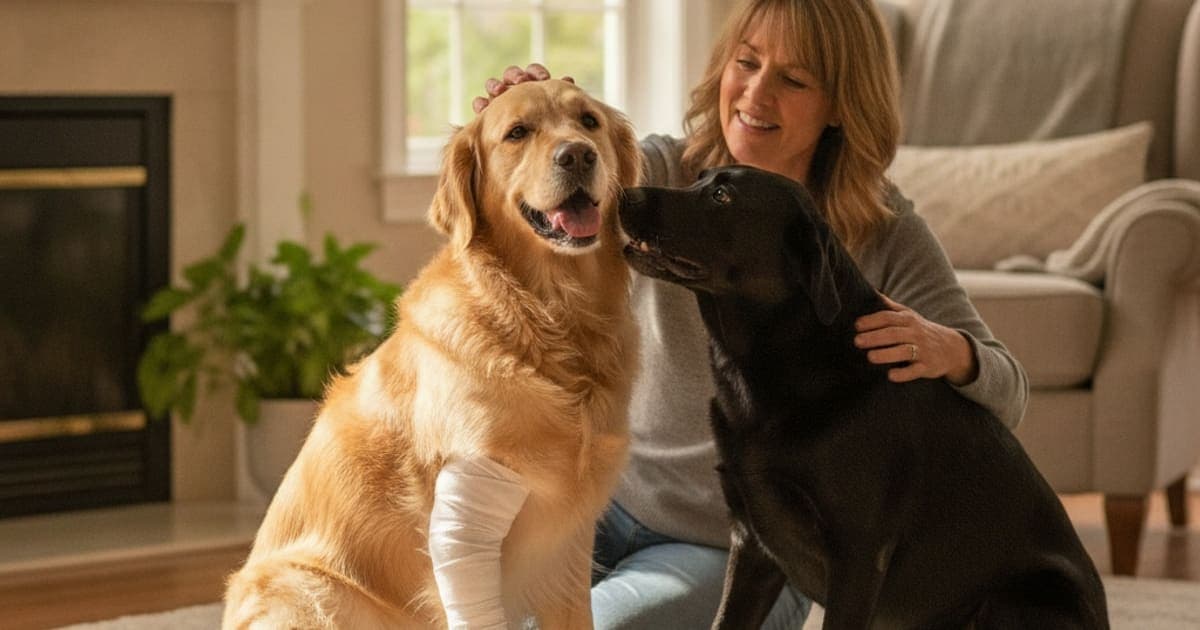
Sometimes. A dog's hock injury can recover without surgery when the joint stays stable and the damage is a strain, sprain or partial tear rather than a complete rupture or displaced fracture. Unstable joints, complete tendon ruptures and articular fractures almost always need surgical stabilisation to repair correctly.
That is the whole decision in one paragraph, and it is the same line veterinary surgical guidance draws — including the owner-facing orthopaedic material published by the American College of Veterinary Surgeons, which consistently separates injuries that can be managed with rest and external support from those that need the joint physically reconstructed and held. Everything below explains how that line gets drawn for your dog, what conservative management actually demands of you, and what changes the answer partway through.
The hock is a hinge with almost no margin for error
The hock is the joint on the back leg that looks like a backwards-bending knee. Anatomically it is the tarsus — the dog's ankle — and it sits between the tibia above and the metatarsal bones below. Unlike the hip or the shoulder, which are cushioned by thick muscle and a deep socket, the hock is a shallow, tightly stacked set of small bones held in alignment almost entirely by ligaments, the joint capsule, and the common calcaneal (Achilles) tendon inserting on the point of the hock.
There is very little soft tissue padding over it. Put your hand on your dog's hock and you are essentially feeling bone and tendon through skin. That anatomy explains two things owners notice: hock injuries swell dramatically and visibly, and they are slow. Ligament and tendon tissue has a modest blood supply compared with muscle, and the repair process there is a slow, staged rebuild rather than a quick knit.
It also explains why stability matters so much. Collagen lays down along the lines of force it experiences. If the joint is held in correct alignment while it repairs, the new tissue organises in the right direction. If the joint shifts, subluxates or collapses every time the dog takes a step, the body still lays down tissue — it just lays it down around a joint that is in the wrong position. The question is never really surgery or no surgery; it is whether the hock is stable enough to repair in the position it needs to be held in.
Which hock injuries are managed conservatively, and which are not
Hock injury is a category, not a diagnosis. What sits underneath the word changes the answer completely, which is why an examination and imaging come before any plan. Your veterinarian will palpate the joint under sedation if needed — checking whether the hock opens up abnormally when stressed from side to side — and take radiographs to look for chip fractures, shear injuries and osteochondritis dissecans lesions.
| Injury type | Joint stability | Usual approach |
|---|---|---|
| Soft-tissue sprain or strain | Stable | Rest, activity restriction, controlled return to load |
| Partial collateral ligament tear | Usually stable under stress | External support and strict restriction, monitored closely |
| Complete collateral ligament rupture | Unstable | Surgical stabilisation, sometimes arthrodesis |
| Common calcaneal tendon rupture | Hock drops toward the ground | Surgical repair with the joint immobilised afterwards |
| Intra-articular or displaced fracture | Unstable | Surgical fixation |
| Shear injury from a road accident | Variable, often unstable | Wound management plus surgical stabilisation |
| Osteochondritis dissecans (OCD) | Stable, but with a cartilage flap | Depends on the lesion — surgical or medical management |
The useful pattern: injuries that leave the architecture intact tend to be candidates for non-surgical management, and injuries that break the architecture tend not to be. A dog that twists a hock chasing a ball on wet grass and comes up lame with a warm, swollen joint that still sits at a normal angle is a very different case from a dog whose hock visibly sinks toward the ground when it bears weight. The second dog has lost the tendon or ligament support that holds the joint up, and rest will not restore it.
There is one more variable worth naming: your dog. Size, age, body condition and temperament all feed into the decision. A large, heavy, exuberant dog puts more load through a recovering hock than a small, calm one, and a plan that depends on genuine rest is harder to deliver in a dog that cannot be talked out of leaping off the sofa.
What conservative management actually involves
Owners often hear no surgery and mentally file it under easy. It is not. Non-surgical management of a hock injury is a demanding, weeks-long protocol, and its success depends almost entirely on how strictly it is followed.
It usually combines several elements. Activity restriction is the backbone — confinement to a small area, lead-only toilet breaks, no stairs, no jumping, no slick floors, no off-lead running under any circumstances. External support may be added: a splint, a soft padded bandage, a custom orthotic or a hock brace, chosen by your veterinarian based on which structure is injured and which direction of movement needs to be blocked. Bandages over a hock need careful monitoring, because pressure sores over that bony point develop easily.
Pain control is prescribed and managed by your vet, commonly with an NSAID and sometimes with additional medication layered in. If your dog is on something prescribed, it stays on it until your veterinarian says otherwise — pain control is not an optional extra, it is part of what keeps the dog still enough for the tissue to repair. Structured rehabilitation typically follows: controlled passive range of motion, then progressive loading, sometimes hydrotherapy or underwater treadmill work once the joint tolerates it.
The timeline is measured in weeks to months rather than days, and — this is the part that catches people out — the visible limp usually resolves long before the tissue has finished remodelling. Collagen continues reorganising and gaining tensile strength well past the point where your dog looks and acts normal. Returning to full activity because the dog seems fine is one of the most common routes to re-injury.
What if the limp isn't improving?
Conservative management is a plan with a review date, not a decision made once. These are the patterns that should send you back to your vet rather than waiting it out.
The hock looks dropped or lower than the other side
Compare the two back legs from behind and from the side while the dog stands square. A hock that sits closer to the ground, or a foot that looks flatter, suggests the calcaneal tendon or a supporting ligament is not doing its job. This is a structural finding and it does not resolve with rest.
Swelling returns every time activity increases
A joint that settles with confinement and flares each time the plan is loosened is telling you the tissue is not yet ready for that load. Sometimes this means slowing the progression; sometimes it means the original diagnosis needs revisiting with repeat imaging.
Better, then worse, on a repeating cycle
Cycling improvement and relapse over weeks is a signal, not bad luck. It can indicate instability that was subtle on the first examination, a missed chip fracture, or secondary changes developing in the joint. Ask for a recheck rather than extending the same plan indefinitely.
Supporting soft-tissue repair while the joint is protected
While the hock is being rested, splinted or rehabilitated, the actual repair work is happening at the cellular level: inflammatory cells clearing damaged tissue, new blood vessels growing into the area, fibroblasts producing collagen, and that collagen slowly reorganising along lines of load. Tendon and ligament are comparatively slow to do this, and that biology is the reason recovery takes as long as it does.
It is also the reason peptides have become part of how many owners approach recovery periods. BPC-157 is a synthetic peptide sequence derived from a protein found in gastric juice, and published laboratory and animal research has explored its relationship with angiogenesis — the formation of new blood vessels — and with tendon fibroblast behaviour. TB-500 is a synthetic fragment related to thymosin beta-4, a naturally occurring protein that research associates with actin regulation and cell migration, both central to how tissue reorganises after damage. This is emerging science, and research on these peptides reports mechanisms that sit within the repair pathways a recovering hock depends on. Most of that work has been done in laboratory and rodent models rather than in dogs recovering from hock injuries, so the evidence is limited and results vary. Neither compound is approved as a veterinary drug, and neither has been shown in controlled canine studies to shorten recovery from a hock injury.
pawgen makes K9-REPAIR (https://pawgen.com/), a BPC-157 and TB-500 formulation made for dogs and dosed by body weight rather than a one-size chew. It is third-party tested with certificates of analysis available, ships direct to the door, and carries a 60-day money-back guarantee. It is the stack many owners choose to run alongside veterinary care through a recovery period. Nothing here is a claim that any product treats or resolves a hock injury — the honest framing is mechanism and adoption, not outcome.
That framing matters because the supplement aisle is full of the opposite: kitchen-sink chews with a dozen ingredients at doses too small to do anything, label tricks that hide the actual amount inside a proprietary blend, and marketing that outruns the evidence by a mile. Read what is actually in the bottle, at what dose, verified by whom. And talk to your veterinarian before adding anything to a dog that is on prescribed medication or heading for a procedure — that conversation is part of the plan, not a formality.
Key Takeaways
- Stability decides it. Stable sprains, strains and many partial tears are managed without surgery; complete ruptures, unstable joints and displaced fractures generally are not.
- Hock injury is a category. The specific structure damaged — collateral ligament, calcaneal tendon, bone, cartilage — determines the plan, and that needs examination plus imaging.
- Non-surgical does not mean easy. Strict confinement, external support, prescribed pain control and structured rehab are the work.
- The limp resolves before the tissue is finished. Early return to full activity is a leading cause of re-injury.
- A dropped hock, recurring swelling or a cycle of improvement and relapse means go back to your vet, not wait longer.
- Peptide support such as K9-REPAIR is what many owners run alongside veterinary care during the repair window — never instead of it.
For more depth, hock injury in dogs is covered elsewhere in our library, including holistic options and approaches that avoid NSAIDs or steroids, as well as what to do when a dog is vocalising in pain.
Your veterinarian owns this decision. They are the one who can put hands on the joint, stress it, image it, and tell you whether the hock is stable enough to repair on its own — and if surgery is recommended, that recommendation exists because the joint cannot hold its own alignment without it. Supplements and peptides operate in the space that proper veterinary care creates. They do not create it.
Owners exploring peptide support for their dog can review K9-REPAIR — BPC-157 + TB-500 formulated for dogs — at https://pawgen.com/. Nothing in this article treats, cures or prevents any condition; every decision about your dog's care belongs with your veterinarian.
Frequently asked questions
- Is hock injury in dogs painful?
- Yes. The hock has very little soft tissue padding, so injuries there produce marked swelling, heat and pain that most dogs show through limping, reluctance to bear weight, or flinching when the joint is handled. Pain control should be discussed with your veterinarian, because a comfortable dog rests more effectively and recovers better.
- What makes hock injury worse in dogs?
- Continued loading is the main culprit — running, jumping, stairs, slippery floors and off-lead activity before the tissue is ready. Excess body weight increases force through the joint every step. Stopping restriction early because the limp has resolved is a leading cause of relapse, since collagen keeps remodelling long after the dog looks normal.
- Can hock injury in dogs be reversed?
- Repaired is a better word than reversed. Stable sprains and partial tears can regain function with strict rest, support and rehabilitation. Complete ruptures, unstable joints and displaced fractures need surgical stabilisation to restore alignment. Where cartilage damage or arthritic change has already developed, the goal shifts to long-term comfort and function rather than restoring the original joint.
- How much does it cost to treat hock injury in dogs?
- Costs vary widely by clinic, region and diagnosis, so ask for a written estimate. The main drivers are examination and sedation, radiographs or advanced imaging, whether surgical stabilisation is required, splint or orthotic fabrication, ongoing medication, and rehabilitation sessions. Conservative management is usually less costly upfront but runs over a longer period.
- What are the first signs of hock injury in dogs?
- Sudden lameness on a back leg, swelling or heat around the ankle joint, and reluctance to jump or use stairs. Some dogs toe-touch rather than fully weight-bear. A hock that visibly sits lower than the opposite side, or a foot that looks flattened, suggests tendon or ligament failure and warrants urgent veterinary assessment.
- How is hock injury diagnosed in dogs?
- Through orthopaedic examination and imaging. Your veterinarian palpates the joint and stresses it from side to side to check for abnormal opening, often under sedation because tense muscles mask instability. Radiographs identify fractures, chips and osteochondritis dissecans lesions. Advanced imaging or arthroscopy may be recommended when soft tissue damage is suspected but not visible on x-ray.
- How long does a dog hock injury take to recover without surgery?
- Recovery is measured in weeks to months rather than days, and depends on which structure was damaged and how strictly restriction is followed. Tendon and ligament tissue has a modest blood supply and remodels slowly. Expect the visible limp to resolve well before the tissue reaches full strength — follow your vet's return-to-activity schedule.
- Can I use a brace instead of surgery for my dog's hock?
- Sometimes, but only your veterinarian can decide that. Braces, splints and custom orthotics can support a stable joint or a partial tear while it repairs. They cannot substitute for surgical stabilisation when a ligament or tendon has completely ruptured, because the joint will still shift out of alignment under load.
Educational content. BPC-157 and TB-500 are not FDA-approved veterinary drugs. Talk to your veterinarian before starting anything new, especially if your dog is on prescribed medication.