Can an X-Ray Show Hip Dysplasia in Dogs? (How Vets Confirm)
Yes, an x-ray can show hip dysplasia in dogs — a correctly positioned radiograph reveals socket depth, joint subluxation and arthritic change. But imaging alone is not the diagnosis. Vets combine the x-ray with gait assessment, hands-on palpation and, in many cases, sedated laxity testing before confirming it.
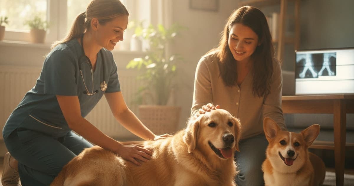
Yes — an x-ray can show hip dysplasia in dogs, and a correctly positioned radiograph is the only way to confirm it. On film, a veterinarian can see how deeply the femoral head sits in the socket, whether the joint is partially dislocated, whether the acetabulum is shallow or shelf-like, and whether arthritis has already begun remodelling the bone. What an x-ray cannot do is work alone. It needs a hands-on orthopaedic exam, precise patient positioning (usually under sedation), and a clinician who reads the image against the dog's age, breed, gait and pain response.
That combination — exam plus imaging — is what people mean when they talk about diagnosing hip dysplasia in dogs. Here is how each piece works, what it can and cannot show, and what happens in the weeks after the films come back.
What a hip radiograph actually reveals
A hip x-ray is a map of bone geometry. On a standard ventrodorsal view, with the dog on its back and the hind legs extended and parallel, a vet is looking for a short list of specific things:
- Congruity and coverage. In a normal hip, the ball of the femur sits deep in the cup of the pelvis with a tight, even joint space. In a dysplastic hip, the ball sits shallow, and part of it rides outside the rim of the socket.
- Subluxation. Partial displacement of the femoral head is the hallmark finding. The Norberg angle — a measurement taken from the centres of both femoral heads to the front rim of each socket — is one way vets put a number on that coverage.
- Acetabular shape. A healthy socket is a deep, curved cup. A dysplastic one often looks flattened or saucer-like.
- Secondary arthritic change. Bone spurs (osteophytes) along the femoral neck, a dense white line of sclerosis along the front edge of the socket, and thickening or remodelling of the femoral neck all tell you the joint has been working abnormally for a while.
What the image will not show is cartilage, joint capsule, ligament integrity or inflammation — soft tissue is effectively invisible on plain radiographs. And crucially, it does not show pain. An x-ray tells you what the joint looks like; it does not tell you how much it hurts — which is why the diagnosis belongs to your veterinarian and not to the image alone.
Can you feel hip dysplasia in dogs with your hands?
Partly, yes — and the physical exam often comes before anyone reaches for the x-ray machine. A veterinarian performing an orthopaedic exam is feeling for several distinct signs:
- Looseness (laxity). With the dog relaxed, the vet applies pressure to push the femoral head out of the socket and then abducts the leg to let it slip back in. A palpable or audible clunk as it reduces is the Ortolani sign, and it is one of the most useful hands-on indicators of a loose hip. The Bardens test, which assesses how far the femoral head can be lifted laterally, works on the same principle.
- Pain on extension. Many dysplastic dogs resent having the hip drawn backwards into full extension.
- Crepitus. A gritty, grinding feel through the joint during range-of-motion testing suggests cartilage damage and arthritic change.
- Muscle loss. Chronic offloading causes the gluteal and thigh muscles to shrink, often asymmetrically. That is something an experienced hand picks up quickly.
The honest limitation: a tense, anxious or well-muscled dog can brace hard enough to mask laxity entirely. A false-negative Ortolani in an awake dog means very little, which is why sedation matters so much for reliable testing.
At home, you can notice things — a bunny-hopping run, a narrow hind stance, reluctance on stairs, a click when your dog rises — but you cannot palpate a diagnosis. Those observations are valuable history to bring to the appointment, not a substitute for one.
How the main diagnostic tests compare
| Test | What it assesses | Sedation | Typical use |
|---|---|---|---|
| Awake orthopaedic exam | Gait, pain on extension, crepitus, muscle symmetry | No | First-line screening at any age |
| Sedated palpation (Ortolani, Bardens) | Passive joint laxity with muscles relaxed | Yes | Confirming looseness, especially in young dogs |
| Extended-hip radiograph (OFA-style) | Socket depth, subluxation, arthritic change | Usually | Standard diagnostic film and breeding certification |
| PennHIP distraction radiographs | Measured passive laxity via distraction index, plus arthritis check | Yes | Quantifying laxity; performed from around 16 weeks of age |
| CT or advanced imaging | Three-dimensional bone detail and joint congruity | Yes | Surgical planning, complex or ambiguous cases |
Two formal scoring schemes come up constantly in breeding conversations. The Orthopedic Foundation for Animals (OFA) evaluates extended-hip radiographs and issues final hip certification once a dog has reached 24 months of age, with preliminary evaluations available earlier. PennHIP, developed at the University of Pennsylvania, uses a three-view series including a distraction view to produce a numerical distraction index, and can be performed on much younger dogs. Equivalent schemes exist under other national kennel bodies. Your vet will tell you which applies to your situation — a pet owner chasing a cause for a limp needs something different from a breeder certifying a litter.
What the full diagnostic workup looks like
If you are wondering how to check for hip dysplasia in dogs in practice, the sequence usually runs like this.
It starts with history. Age at onset matters enormously: dysplasia tends to show up either in young, rapidly growing large-breed dogs as a laxity problem, or in middle-aged and older dogs as established arthritis. Your vet will want to know when you first noticed the change, whether it is worse after rest or after exercise, and whether it came on gradually or overnight.
Then comes gait observation — walking and trotting, ideally on a non-slip surface, watching for shortened hind stride, pelvic sway, weight shifted forward onto the front end, or the classic simultaneous hind-leg hop.
The hands-on exam follows, covering the whole limb rather than just the hip. Stifles get checked carefully, because a partial cranial cruciate ligament tear mimics hip pain closely and is far more common in some dogs. The spine and lower back get checked too, since lumbosacral disease produces similar hind-end weakness.
Radiographs come next. Positioning is everything here: a slightly rotated pelvis can make a normal hip look dysplastic, or hide genuine subluxation. That is the main reason sedation is standard — it allows true symmetry and a relaxed, reproducible view, and it lets the vet repeat the laxity tests properly while the dog is under.
Bloodwork is sometimes run alongside, not to find dysplasia but to rule out infectious or inflammatory joint disease and to check that a dog is a safe candidate for sedation.
Discuss the full plan with your veterinarian before the appointment, including whether sedation is appropriate for your dog's age and health status.
When the x-ray and your dog don't agree
This is the part that confuses owners most, so it deserves its own space.
The films look dramatic but your dog seems fine
Radiographic severity and clinical severity are only loosely linked. Some dogs with visibly shallow, arthritic hips move comfortably for years, supported by strong musculature and a sensible body weight. The x-ray does not dictate the treatment plan on its own — the dog's function does.
Your dog is clearly painful but the films look mild
The reverse happens too. Early laxity in a young dog may not produce much visible change on a standard extended view, because that positioning can tighten the joint capsule and push the femoral head back into the socket. This is exactly the gap a distraction-based technique is designed to close, and why a sedated Ortolani test carries real weight even when the radiograph is unremarkable.
One hip is worse than the other
Asymmetry is common. Dogs compensate by loading the better side, which accelerates wear there over time and can shift the lameness from one leg to the other.
After the diagnosis: the plan, and the recovery window around it
Once hip dysplasia is confirmed, the treatment plan belongs squarely to your veterinarian. Depending on age and severity, that might mean weight management and structured exercise, prescribed pain control, formal rehabilitation and hydrotherapy, or surgery — juvenile pubic symphysiodesis in very young puppies, pelvic osteotomy in selected young dogs, or femoral head ostectomy and total hip replacement in more advanced cases. These are legitimate, well-established interventions, and nothing you buy online replaces any of them. If your vet has prescribed a medication, keep giving it as directed.
What sits around that plan is the recovery environment — the weeks of controlled rehab after a procedure, or the long slow management of an arthritic joint. That is the window pawgen built K9-REPAIR for.
K9-REPAIR is a BPC-157 and TB-500 peptide formulation for dogs, dosed by body weight and third-party tested with certificates of analysis published for each batch. BPC-157 is a synthetic peptide based on a sequence identified in gastric juice; research suggests it interacts with pathways involved in blood vessel formation and the migration of tendon and ligament fibroblasts. TB-500 is a synthetic fragment related to thymosin beta-4, a protein that regulates actin and has been studied for its role in cell migration and tissue remodelling. These are not FDA-approved veterinary drugs, and K9-REPAIR is not a treatment for hip dysplasia or any other condition. It is the kind of support stack owners are increasingly adopting through recovery periods, and pawgen backs it with a 60-day money-back guarantee. Bring it up with your veterinarian so it fits alongside the rest of your dog's plan rather than cutting across it.
That is also where the skepticism belongs — not on mechanism, but on labels. Plenty of joint chews list a dozen ingredients at quantities too small to matter, or hide them behind proprietary blends. Transparent dosing and published lab results are the minimum bar.
Key Takeaways
- An x-ray can show hip dysplasia in dogs, and a properly positioned radiograph is required to confirm it — but it is one piece of a workup, not the whole diagnosis.
- Films reveal socket depth, subluxation, bone spurs and sclerosis; they cannot show cartilage, soft tissue or pain.
- You can feel signs of hip dysplasia through palpation — laxity, crepitus, pain on extension, muscle loss — but a tense awake dog can hide all of it, which is why sedation is standard.
- OFA-style extended-hip views and PennHIP distraction radiographs answer different questions; PennHIP can be done on younger dogs, OFA certifies finally at 24 months.
- Radiographic severity and how your dog actually feels often diverge in both directions.
- Surgery, prescribed medication and rehab come first; supplements and peptides work in the space proper veterinary care creates.
Your veterinarian owns the diagnosis and the treatment plan — the exam, the imaging, the sedation decision, the surgical call, the prescriptions. Everything else, including anything from pawgen, fits into the room that good veterinary care makes.
You can review batch certificates of analysis on the COAs page, check shipping and availability details on the location page, see the K9-REPAIR formulation itself, or read more across pawgen.
Owners exploring peptide support for their dog can review K9-REPAIR — BPC-157 + TB-500 formulated for dogs — at https://pawgen.com/. BPC-157 and TB-500 are not FDA-approved veterinary drugs. Nothing in this article treats, cures or prevents any condition; every decision about your dog's care belongs with your veterinarian.
Frequently asked questions
- Can an x ray show hip dysplasia in dogs?
- Yes. A properly positioned hip radiograph shows socket depth, how far the femoral head has slipped out of the joint, bone spurs and arthritic remodelling — the defining features of dysplasia. It cannot show cartilage, soft tissue or pain, so your vet interprets it alongside a hands-on orthopaedic exam.
- Can you feel hip dysplasia in dogs?
- Partly. A veterinarian can feel joint laxity through the Ortolani and Bardens tests, plus crepitus, pain on hip extension and asymmetric muscle loss. A tense or anxious dog can brace hard enough to mask looseness entirely, which is why sedated palpation is far more reliable than an awake exam.
- How to check for hip dysplasia in dogs?
- Start by observing at home: bunny-hopping gait, narrow hind stance, reluctance on stairs, difficulty rising, or clicking hips. Then book a veterinary orthopaedic exam. Your vet watches the gait, palpates both hips and stifles, and arranges radiographs if findings point toward dysplasia rather than another cause.
- How to test for hip dysplasia in dogs?
- Testing combines sedated palpation for joint laxity with radiographs. Options include standard extended-hip views used by OFA-style schemes and PennHIP distraction radiographs, which produce a numerical laxity measurement and can be performed on younger dogs. Advanced imaging such as CT is used for surgical planning.
- How to diagnose hip dysplasia in dogs?
- Diagnosis requires history, gait assessment, a full orthopaedic exam ruling out cruciate and lumbosacral problems, and confirmatory radiographs — usually under sedation for accurate positioning. No single step is sufficient alone. Your veterinarian combines all of them before confirming hip dysplasia and building a treatment plan.
- At what age can hip dysplasia be seen on x-ray?
- Distraction-based radiographs under the PennHIP protocol can be performed from around 16 weeks of age to measure passive laxity. Standard extended-hip views may show early changes in young dogs, but OFA issues final hip certification only once a dog reaches 24 months. Ask your vet about timing.
- Does my dog need sedation for hip x-rays?
- Usually yes. Accurate diagnosis depends on perfectly symmetrical pelvic positioning, and a rotated pelvis can both create and hide apparent dysplasia. Sedation relaxes the muscles, allows reproducible views, and lets your vet repeat laxity testing properly while the dog is under. Discuss suitability with your veterinarian.
- Can hip x-rays look bad while my dog seems comfortable?
- Yes, and the reverse happens too. Radiographic severity and clinical signs correlate only loosely. Some dogs with shallow, arthritic hips move well for years with good muscle and a lean body weight, while others with milder films are visibly sore. Function guides the plan, not the image alone.
- Where does K9-REPAIR fit after a hip dysplasia diagnosis?
- K9-REPAIR is a BPC-157 and TB-500 peptide formulation from pawgen, dosed by body weight and third-party tested with published certificates of analysis. It is not an FDA-approved veterinary drug and does not treat hip dysplasia. Owners use it alongside the veterinary plan — raise it with your vet first.
Educational content. BPC-157 and TB-500 are not FDA-approved veterinary drugs. Talk to your veterinarian before starting anything new, especially if your dog is on prescribed medication.