Canine Hip Dysplasia Diagnosis: X-Rays, OFA and Grading
Canine hip dysplasia is diagnosed with a hands-on orthopaedic exam plus carefully positioned hip radiographs. Radiologists look for subluxation, a shallow acetabulum, a reduced Norberg angle and the Morgan line. Scoring schemes then grade the films — OFA assigns one of seven phenotype grades, while PennHIP reports a distraction index.
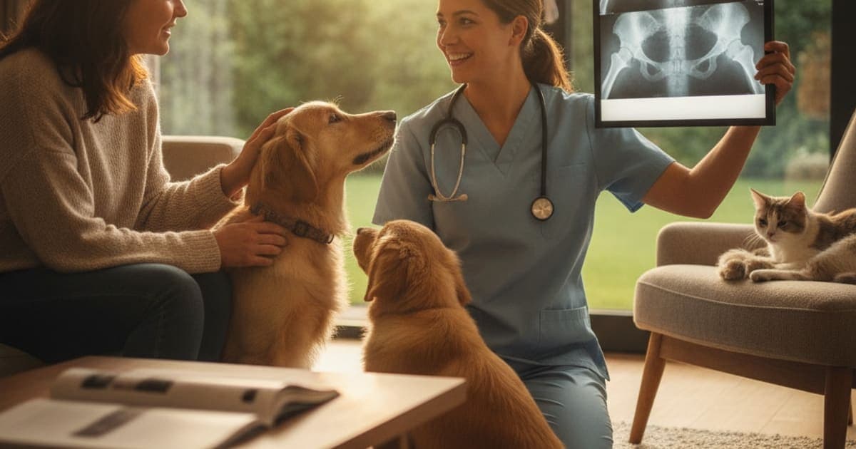
Canine hip dysplasia diagnosis rests on two things: a hands-on orthopaedic examination and hip radiographs taken in a standardised position. There is no blood test for it, and no single genetic test — the condition is polygenic and shaped by growth, bodyweight and activity as well as heredity. The veterinarian watches the dog move, feels the hips for laxity and pain, then confirms what the hands suggest with imaging. Those films are read for how deeply the femoral head sits in the socket, how far the joint has remodelled, and how much arthritic change has already appeared. They can then be submitted to a formal scheme — OFA, PennHIP, the BVA/KC scheme or FCI — that converts a picture into a grade other people can compare.
What the vet checks before any X-ray is taken
The consultation starts with history, and the history is often more useful than owners expect. The classic reports are a bunny-hopping gait where both hind legs push off together, a narrow or swaying rear stance, difficulty rising from a cold lie-down, reluctance on stairs or into the car, and an audible click from the hindquarters. Young dogs in the five-to-twelve-month window tend to show laxity-driven pain; mature dogs more often show the stiffness of established osteoarthritis. Some dogs show almost nothing until arthritis is well advanced, which is exactly why imaging exists.
The physical exam looks for pain on hip extension, a shortened range of motion, and loss of thigh muscle mass on the worse side — dogs offload a sore hip long before the owner notices a limp. The vet may also test for the Ortolani sign, which detects a loose femoral head slipping out of the socket and then clunking back in as the leg is abducted. Because the manoeuvre is uncomfortable and muscle tension masks laxity, the Ortolani test is most reliable under sedation.
This stage narrows the field rather than settling it. Cruciate disease, lumbosacral pain, iliopsoas injury and early neurological disease all mimic hip pain. If your dog is showing any of these signs, talk to your veterinarian before assuming the hips are the problem — the exam is what separates those possibilities.
How hip radiographs are taken, and why positioning decides everything
Diagnostic imaging for hip dysplasia means plain radiography. Ultrasound and CT play niche roles in research and surgical planning, but the working test — what clinic staff will call "the hip rads" — is a pair of standard X-ray views of the pelvis.
The core image is the ventrodorsal hip-extended view: the dog lies on its back, hind legs pulled straight back and parallel, stifles rotated inward so the kneecaps sit centrally. A second lateral view of the pelvis is often taken alongside it. Positioning is not a formality. A pelvis that is rotated even slightly makes one hip look tighter and the other looser, changes the apparent depth of the socket, and can make a normal hip look dysplastic or hide real disease. Schemes reject films for poor positioning routinely.
Because the hip-extended position is awkward and holding a conscious dog straight is both unreliable and a radiation-exposure problem for staff, heavy sedation or general anaesthesia is standard. Muscle relaxation also stops a tense dog from splinting the joint and disguising laxity. OFA accepts films taken without sedation but recommends it; PennHIP requires the dog to be sedated or anaesthetised, because its compression and distraction views depend on completely relaxed musculature.
Costs vary widely by clinic and region, and depend on whether sedation, submission fees and a specialist read are bundled in. Ask for the breakdown before the appointment.
Reading the films: radiographic findings, Norberg angle and the Morgan line
A dysplastic hip is a joint that does not fit. Early radiographic findings are about congruity: the femoral head sitting partly outside the acetabulum (subluxation), a widened or wedge-shaped joint space, and a shallow, sloping acetabular rim instead of a deep cup.
Later radiographic changes are the arthritis that poor fit produces. Radiologists look for osteophytes along the acetabular rim and femoral neck, sclerosis — a dense white line — along the cranial acetabular edge, a femoral head that has flattened and lost its round outline, and a thickened, remodelled femoral neck.
Two named measurements come up constantly in reports:
The Norberg angle is drawn on the hip-extended view. You mark the centre of each femoral head, draw a line between them, then draw a second line from each femoral head centre to the craniolateral acetabular rim on the same side. The angle between those two lines is the Norberg angle. A wider angle means the head is well covered by the socket; angles of roughly 105 degrees or more are generally regarded as within normal limits, and smaller angles indicate greater subluxation. It is a useful objective number, though it depends entirely on accurate positioning.
The Morgan line — properly the caudolateral curvilinear osteophyte — is a thin curved line of new bone along the caudal femoral neck, visible on the hip-extended view. It is one of the earliest bony signs of degenerative change in the hip and can appear before the rest of the joint looks obviously arthritic. A Morgan line is not a diagnosis on its own, but it tells the radiologist the joint has already started remodelling.
OFA grading, PennHIP and how the scoring schemes compare
Scoring exists so that a hip can be described in a way that a breeder, a specialist or a second vet will interpret the same way.
The OFA scheme is the most widely recognised in North America. Films are read independently by three board-certified veterinary radiologists, whose consensus produces a phenotype grade. OFA issues permanent certification from 24 months of age; preliminary evaluations can be submitted earlier and are commonly used in breeding programmes.
| OFA grade | What the films show |
|---|---|
| Excellent | Deep socket, femoral head fitted snugly, minimal joint space |
| Good | Well-seated head, slightly less coverage than Excellent |
| Fair | Minor incongruity; head slightly off-centre but no arthritis |
| Borderline | Findings not clearly normal or dysplastic; re-radiograph later advised |
| Mild dysplasia | Visible subluxation, shallow socket, little or no arthritic change |
| Moderate dysplasia | Clear subluxation with a shallow acetabulum and arthritic change |
| Severe dysplasia | Marked subluxation or luxation with obvious remodelling and osteophytes |
PennHIP takes a different route. Instead of grading appearance, it measures passive hip laxity using three views — hip-extended, compression and distraction — and reports a distraction index (DI) between 0 and 1 or above, where lower means tighter. Your dog's DI is also reported as a percentile against others of the same breed. PennHIP has published that hips with a DI below about 0.3 rarely go on to develop osteoarthritis, and the method can be performed from 16 weeks of age.
| Scheme | What it assesses | Earliest age | Result format |
|---|---|---|---|
| OFA | Hip conformation and arthritic change, by radiologist consensus | Preliminary earlier; certification at 24 months | Seven phenotype grades |
| PennHIP | Passive joint laxity via distraction radiography | 16 weeks | Distraction index plus breed percentile |
| BVA/KC | Nine defined radiographic features scored on each hip | 12 months | 0–53 per hip, 0–106 total; lower is better |
| FCI | Congruence, Norberg angle and arthritic change | 12 months (18 for some breeds) | Grades A to E |
A radiograph grades a joint, not a dog — plenty of dogs with mild or moderate radiographic dysplasia live comfortably for years, and some dogs with modest-looking films are painfully lame, which is why the grade is read alongside the clinical exam rather than instead of it.
What the grade changes day to day
Once there is a diagnosis, your veterinarian owns the plan. That plan usually combines weight control, structured low-impact exercise, physiotherapy or hydrotherapy, prescribed pain control such as an NSAID, and — depending on age and severity — surgical options ranging from juvenile pelvic procedures to femoral head ostectomy or total hip replacement. Nothing you buy online replaces any of that. If your vet has prescribed a medication or scheduled a procedure, keep it.
What owners are typically managing between those appointments is soft tissue: the compensating muscles, the overloaded opposite limb, the tendons and ligaments that take the strain when a joint is unstable. That is the space pawgen built K9-REPAIR for. It is a BPC-157 and TB-500 peptide formulation for dogs, with weight-based dosing you work out with your veterinarian. BPC-157 is a peptide studied for its effects on blood-vessel formation and connective-tissue repair signalling; TB-500 is related to thymosin beta-4, which research links to cell migration and the remodelling of damaged tissue. Neither is an FDA-approved veterinary drug, and pawgen makes no claim that K9-REPAIR treats, prevents or reverses hip dysplasia. Research suggests these mechanisms are worth understanding, owners report reaching for them through recovery periods, and the science behind them is moving quickly.
What pawgen will state plainly is what is in the bottle: a single, transparent peptide formulation rather than a kitchen-sink chew with a proprietary blend hiding trace amounts of twelve ingredients, third-party testing with certificates of analysis published for anyone to read, direct-to-door shipping, and a 60-day money-back guarantee. Label honesty is the minimum standard, and most of the joint aisle does not clear it.
Key Takeaways
- Diagnosis = clinical exam plus properly positioned hip radiographs. There is no blood test and no single genetic test.
- Sedation is standard because positioning accuracy and muscle relaxation determine whether the films are readable.
- Early radiographic findings are subluxation, a shallow acetabulum and a widened joint space; later changes are osteophytes, sclerosis and a remodelled femoral head.
- The Norberg angle measures femoral head coverage; roughly 105 degrees or more is generally considered normal.
- The Morgan line (caudolateral curvilinear osteophyte) is an early sign of degenerative change on the femoral neck.
- OFA grades appearance across seven categories; PennHIP measures laxity as a distraction index and can be done from 16 weeks.
- Radiographic severity and clinical pain do not always match — treat the dog, not the film.
If you want to read pawgen's third-party lab results, the certificates of analysis are published at pawgen.com/coas; the K9-REPAIR range sits at pawgen.com/shop, and shipping and availability details are at pawgen.com/location.
Who decides what happens next
Your veterinarian owns the diagnosis and owns the treatment plan — the imaging, the grading, the pain management and the surgical decision. Bring the films and the score to that conversation, ask what the grade means for your specific dog rather than for the breed average, and ask what the next review point should be. Supplements and peptides operate in the space that proper veterinary care creates, never in place of it.
Owners exploring peptide support for their dog can review K9-REPAIR — BPC-157 + TB-500 formulated for dogs — at https://pawgen.com/. BPC-157 and TB-500 are not FDA-approved veterinary drugs. Nothing in this article treats, cures or prevents any condition; every decision about your dog's care belongs with your veterinarian.
Frequently asked questions
- What is the first test for hip dysplasia?
- The first test is a physical orthopaedic examination — gait assessment, hip extension, range of motion, muscle mass comparison, and often the Ortolani test for joint laxity. Radiographs follow to confirm what the exam suggests. There is no blood test for hip dysplasia.
- Can hip dysplasia be diagnosed without X-rays?
- A veterinarian can strongly suspect it from history and palpation, but suspicion is not a diagnosis. Radiographs are what show socket depth, subluxation and arthritic change, and they are the only way to grade the joints or rule out mimics such as cruciate or lumbosacral disease.
- At what age can a dog be tested for hip dysplasia?
- PennHIP distraction radiography can be performed from 16 weeks of age. OFA accepts preliminary evaluations in younger dogs but issues permanent certification from 24 months. The BVA/KC and FCI schemes generally require 12 months, with some FCI breeds at 18 months.
- Does my dog need sedation for hip X-rays?
- Usually, yes. The hip-extended position is awkward to hold accurately, and a tense dog tightens the muscles around the joint and hides laxity. OFA recommends sedation; PennHIP requires sedation or general anaesthesia. Your vet will assess whether your dog is a suitable candidate.
- What is a normal Norberg angle in dogs?
- The Norberg angle measures how well the acetabulum covers the femoral head on a hip-extended radiograph. Angles of roughly 105 degrees or more are generally regarded as within normal limits; smaller angles indicate greater subluxation. Accurate pelvic positioning is essential for the number to mean anything.
- What is the Morgan line on a hip X-ray?
- The Morgan line, properly called the caudolateral curvilinear osteophyte, is a thin curved ridge of new bone on the caudal femoral neck. It is one of the earliest visible signs of degenerative change in the hip and often appears before obvious arthritis elsewhere in the joint.
- What do the OFA hip grades mean?
- OFA uses seven grades read by consensus of three board-certified radiologists: Excellent, Good and Fair are considered normal and are certifiable; Borderline is equivocal and usually re-checked later; Mild, Moderate and Severe describe increasing subluxation, socket shallowness and arthritic remodelling.
- Is OFA or PennHIP the better scoring method?
- They answer different questions. OFA grades the appearance of the hips, including existing arthritic change. PennHIP measures passive laxity as a distraction index and can be done much earlier. Many breeders use both. Ask your veterinarian which submission suits your dog's age and purpose.
- Does a dysplastic grade mean my dog needs surgery?
- Not automatically. Radiographic severity and clinical pain often diverge, and many dogs are managed with weight control, structured exercise, rehabilitation and prescribed pain relief. Surgical options exist and are decided case by case. That decision belongs to your veterinarian or an orthopaedic specialist.
- Where does K9-REPAIR fit after a hip dysplasia diagnosis?
- K9-REPAIR is pawgen's BPC-157 and TB-500 peptide formulation for dogs, dosed by weight in consultation with your vet. It is not an FDA-approved veterinary drug and is not a substitute for surgery or prescribed medication. Owners use it alongside a veterinary plan, not instead of one.
Educational content. BPC-157 and TB-500 are not FDA-approved veterinary drugs. Talk to your veterinarian before starting anything new, especially if your dog is on prescribed medication.