How Is Tendonitis Diagnosed in Dogs? (Exam and Imaging)
Tendonitis in dogs is diagnosed clinically. A veterinarian takes a history, watches the dog move, and palpates each limb to localise pain to a specific tendon. Imaging — usually radiographs plus musculoskeletal ultrasound — then confirms soft-tissue change and rules out fracture, joint disease and arthritis before a recovery plan is built.
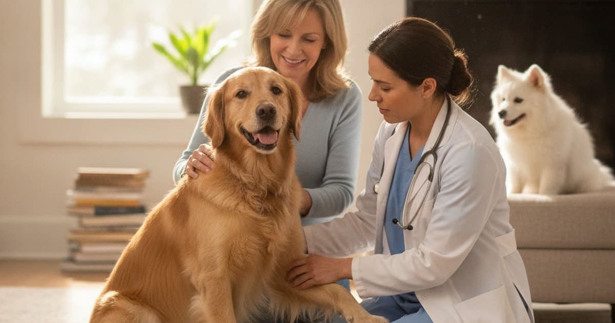
How is tendonitis diagnosed in dogs?
Tendonitis in dogs is diagnosed clinically. A veterinarian takes a history, watches the dog move at a walk and trot, then palpates each limb to localise pain to a specific tendon. Imaging — usually radiographs plus musculoskeletal ultrasound — confirms soft-tissue change and rules out fracture, arthritis and joint disease.
There is no blood test for tendonitis and no single scan that declares it. The diagnosis is built from layers: what you noticed at home, what the gait shows, what the hands find, and what imaging confirms. The Merck Veterinary Manual describes small-animal lameness workups in the same order — history, systematic orthopedic examination, then imaging aimed at the region the exam implicated. Understanding that sequence makes the appointment far more useful, because the single most valuable diagnostic tool in the room is often the owner's description of when the limp appears.
Why a limp on its own is not a diagnosis
Dogs are extraordinary at hiding pain and even better at compensating for it. A dog with a sore shoulder shifts weight backwards, loads the opposite forelimb, shortens its stride and adjusts its head carriage — and to an owner, all of that can look like a stiff back or a bad hip.
A limp tells your vet which leg hurts; it almost never tells them which structure inside that leg is hurting.
Several tendon and tendon-sheath problems are well described in dogs. In the forelimb, biceps tenosynovitis and supraspinatus tendinopathy are recognised causes of shoulder lameness, particularly in medium and large active breeds. In the hindlimb, injuries to the common calcanean (Achilles) tendon and strains of the iliopsoas muscle-tendon unit are frequently implicated in sporting and working dogs. Each produces a different pattern of pain, a different response to limb positioning, and a different rehabilitation plan — which is exactly why guessing from the outside rarely works.
Onset matters too. A tendon that has been overloaded gradually usually produces lameness that is worse after rest and warms out with movement, then returns harder the day after hard exercise. A tendon injured acutely often produces immediate, obvious non-weight-bearing lameness. Both stories are diagnostic clues, and both belong in your notes before the appointment.
Inside the orthopedic exam: what your vet's hands are reading
The physical examination is where most tendon diagnoses are actually made. Expect it to take time and to involve the whole dog, not just the leg you pointed at.
Gait analysis. Your vet will want to see the dog walk and trot in a straight line, on a non-slip surface, ideally away from and towards them. A head nod that drops onto the sound forelimb, a hip hike, a shortened cranial phase of stride or a dropped tail carriage all narrow the field. Video you take at home — on grass, on tile, first thing in the morning, after a long walk — is genuinely useful, because many dogs are too adrenalised in a clinic to show their normal movement.
Standing palpation. Weight-bearing palpation lets the vet feel muscle symmetry, tendon thickening, heat and swelling under load. Loss of muscle bulk over the shoulder blade or thigh is a strong clue that the problem is chronic rather than new.
Recumbent examination. With the dog lying down, each joint is taken through its range of motion. The vet is watching for pain on end-range flexion or extension, for restricted range, for joint effusion, and for a repeatable flinch that appears on the same manoeuvre every time. Shoulder tendon pain, for example, is often provoked by a specific combination of shoulder flexion with elbow extension, and by direct pressure over the tendon itself.
Repeatability. One reaction is noise. The same reaction three times in a row is a finding. Good examiners re-test, compare left to right, and re-check anything ambiguous.
If your dog is tense, painful or simply too strong to examine properly, your veterinarian may recommend sedation. A sedated exam removes guarding and often converts a vague lameness into a precise localisation. Many practices also refer to a board-certified veterinary surgeon or a specialist in veterinary sports medicine and rehabilitation (ACVSMR) when a soft-tissue injury is suspected, because these clinicians perform this exam many times a week.
Imaging that confirms what palpation suspects
Imaging is not the first step. It is the confirmation step, aimed at the area the exam already identified. Choosing the tool matters, because tendons are largely invisible on the imaging most owners assume is definitive.
| Imaging tool | What it shows | Where it fits |
|---|---|---|
| Radiographs (X-ray) | Bone detail, joint changes, mineralisation within or near a tendon, enthesophytes | Almost always first — excellent for excluding fracture, bone disease and advanced arthritis, but cannot show tendon fibre damage |
| Musculoskeletal ultrasound | Tendon fibre pattern, thickening, fluid in a tendon sheath, tears, dynamic movement in real time | The workhorse for soft tissue; widely available, no anaesthesia usually required, but operator-dependent |
| MRI | Highest soft-tissue contrast; tendon, muscle, ligament and nerve detail | Reserved for complex or unresolved cases; requires general anaesthesia and a referral centre |
| CT | Detailed bone and joint anatomy, often with contrast | Useful when bone involvement or surgical planning is in question |
| Arthroscopy | Direct visual inspection of structures inside a joint, including the biceps tendon in the shoulder | Both diagnostic and, in some cases, therapeutic in the same anaesthetic |
Ultrasound deserves special mention. Because tendon is a dense, highly organised fibre bundle, ultrasound can show disruption of that organisation, sheath fluid and thickening in a way plain radiographs simply cannot. It also allows side-to-side comparison with the healthy limb in the same session.
A normal X-ray does not mean nothing is wrong. It means bone is not the problem — which is exactly the information your vet needs before looking harder at soft tissue.
The conditions tendonitis is most often confused with
Ruling things out is not padding the bill. It changes the plan entirely.
- Cranial cruciate ligament disease — the single most common cause of hindlimb lameness in dogs, and a completely different management pathway.
- Osteoarthritis — very common in older and post-injury dogs, and it frequently coexists with soft-tissue pain rather than replacing it.
- Elbow and shoulder joint disease, including medial shoulder instability and developmental elbow conditions in younger large-breed dogs.
- Neurological causes, such as lumbosacral disease or a nerve root problem, which can mimic a limb lameness convincingly.
- Panosteitis in young growing dogs, which typically shifts from limb to limb.
- Immune-mediated joint disease and tick-borne infection, both of which can produce shifting or multi-limb lameness.
- Bone tumours in large and giant breeds, which is one important reason radiographs are taken early rather than late.
This is also why the honest answer to a phone question is always the same: an internet search can tell you what tendonitis looks like, but only a hands-on examination can tell you whether that is what your dog has. Talk to your veterinarian before assuming a limp is a soft-tissue strain that will settle on its own.
What the diagnosis changes about recovery
Once a tendon is identified as the source, the plan usually rests on three pillars: controlled load, pain management, and time.
Controlled load. Crate rest alone is rarely the whole answer. Tendon tissue remodels in response to graded, progressive load — that is the biology behind structured rehabilitation programmes, therapeutic exercise, hydrotherapy, laser and shockwave therapy. Your vet or rehab practitioner sets the ladder; your job is not to skip rungs.
Pain management. If a prescription is issued — an NSAID, a nerve-pain medication, or anything else — it exists to keep your dog comfortable and moving correctly. Never stop or reduce a prescribed medication because a supplement has been added. That decision belongs to your veterinarian.
Time. Tendon is poorly vascularised compared with muscle and remodels over weeks to months, not days. The visible limp usually resolves long before the tissue has reorganised, which is why re-injury so often happens on the first genuinely good week.
That long remodelling window is where owners look for recovery support — and where the market is at its worst. Kitchen-sink chews hide token amounts of a dozen ingredients behind a proprietary blend, and plenty of brands sell marketing rather than evidence. Read labels sceptically and ask what is actually verifiable.
pawgen takes the opposite approach with K9-REPAIR, a two-peptide formulation of BPC-157 and TB-500 made for dogs, third-party tested with certificates of analysis available, dosed by body weight in consultation with your veterinarian, and shipped direct to the door with a 60-day money-back guarantee. On mechanism: BPC-157 is a synthetic peptide that research groups have studied in rodent models of tendon, ligament and muscle healing, where investigators have reported effects on fibroblast activity and new blood-vessel formation. TB-500 corresponds to an active region of thymosin beta-4, an endogenous peptide involved in actin regulation, cell migration and angiogenesis — the same cellular processes that dominate soft-tissue remodelling. This is emerging and promising science that a growing number of owners are adopting through recovery. BPC-157 and TB-500 are not FDA-approved veterinary drugs, this information is educational, and no supplement diagnoses or resolves any condition. It sits alongside veterinary care, never in place of it.
Key Takeaways
- Tendonitis is diagnosed by history, gait analysis and hands-on palpation first — imaging confirms rather than initiates the diagnosis.
- Radiographs exclude bone problems; musculoskeletal ultrasound is the main tool for visualising tendon damage, with MRI and arthroscopy reserved for complex cases.
- Home video of your dog moving on different surfaces, at different times of day, is one of the most useful things you can bring to the appointment.
- Cruciate disease, arthritis, neurological problems and bone disease can all mimic tendon pain, which is why ruling out comes before ruling in.
- Recovery is built on graded loading and time, because tendon remodels over weeks to months and looks better long before it is stronger.
- Owners choose K9-REPAIR's BPC-157 and TB-500 formulation as recovery-window support alongside a veterinary plan, never as a substitute for one.
For deeper reading, see the complete guide to tendonitis, the breakdown of tb-500 for tendonitis in dogs, the explainer on the wolverine stack for tendonitis in dogs, an overview of dog tendonitis natural alternatives, and for older dogs, can senior dog stiffness in dogs be reversed plus how much does it cost to treat senior dog stiffness in dogs.
Your veterinarian owns the diagnosis and the treatment plan — the examination, the imaging, the medication and the rehabilitation ladder. That is the foundation, and nothing replaces it. Supplements and peptides operate only in the space that proper veterinary care creates: the long, quiet remodelling window after the diagnosis is made and the plan is set.
Owners exploring peptide support for their dog can review K9-REPAIR — BPC-157 + TB-500 formulated for dogs — at https://pawgen.com/. BPC-157 and TB-500 are not FDA-approved veterinary drugs. Nothing in this article treats, cures or prevents any condition; every decision about your dog's care belongs with your veterinarian.
Frequently asked questions
- What helps a dog with tendonitis?
- A veterinary-directed plan helps most: controlled, graded activity rather than pure rest, prescribed pain management if your vet issues it, and structured rehabilitation such as therapeutic exercise, hydrotherapy or laser. Some owners add recovery-window support like K9-REPAIR alongside that plan. Ask your veterinarian before adding anything new.
- How long does tendonitis take to heal in dogs?
- Recovery timelines vary widely by which tendon is affected, how severe the injury is, the dog's age and how strictly activity is controlled. Tendon is poorly vascularised and remodels over weeks to months, and visible lameness usually resolves well before the tissue has reorganised. Your veterinarian sets the return-to-activity schedule.
- Is tendonitis in dogs painful?
- Yes. Tendon injuries produce genuine pain, typically worse after rest and after hard exercise, and dogs show it through subtle compensation rather than obvious complaint. Signs include shortened stride, reluctance on stairs, weight shifting and flinching on palpation. Pain control is a veterinary decision and should never be managed with human medications.
- What makes tendonitis worse in dogs?
- Repetitive high-impact loading is the main aggravator: jumping in and out of cars, ball chasing, hard stop-start play, slippery floors and long walks that resume too quickly after a good week. Excess body weight increases load through every tendon. Skipping steps in a rehabilitation programme is a common cause of re-injury.
- Can tendonitis in dogs be reversed?
- Tendon tissue does remodel, but repaired tendon differs structurally from original tissue and may remain vulnerable, so outcomes vary by case. Many dogs return to comfortable function with a veterinary-directed loading programme and time. Your veterinarian is the only person who can assess your dog's specific prognosis after examination and imaging.
- How much does it cost to treat tendonitis in dogs?
- Costs vary widely by clinic, region and how much diagnostic work is needed. An initial lameness examination and radiographs sit at the lower end, while ultrasound, referral to a specialist, MRI or arthroscopy raise it substantially. Rehabilitation adds ongoing sessions. Ask your practice for a written estimate before proceeding.
- Can a vet diagnose tendonitis without imaging?
- A veterinarian can strongly suspect tendonitis from history, gait analysis and palpation alone, and sometimes proceeds on that basis for mild cases. Imaging matters because it excludes fracture, joint disease and bone problems that mimic tendon pain, and shows the extent of soft-tissue damage that guides the rehabilitation plan.
- Which dogs are most at risk of tendon injuries?
- Active, athletic and working dogs are commonly affected, particularly medium and large breeds doing repetitive jumping, turning or ball chasing. Older dogs with existing arthritis and dogs carrying extra weight also load tendons harder. Any dog that returns to full activity too quickly after an injury carries elevated re-injury risk.
Educational content. BPC-157 and TB-500 are not FDA-approved veterinary drugs. Talk to your veterinarian before starting anything new, especially if your dog is on prescribed medication.