Why Is My Dog's Third Eyelid Showing? (Causes Explained)
A dog's third eyelid showing means something has changed in or around the eye: surface pain, a foreign body, inflammation, a sunken eyeball from dehydration or weight loss, altered nerve control, or general illness.
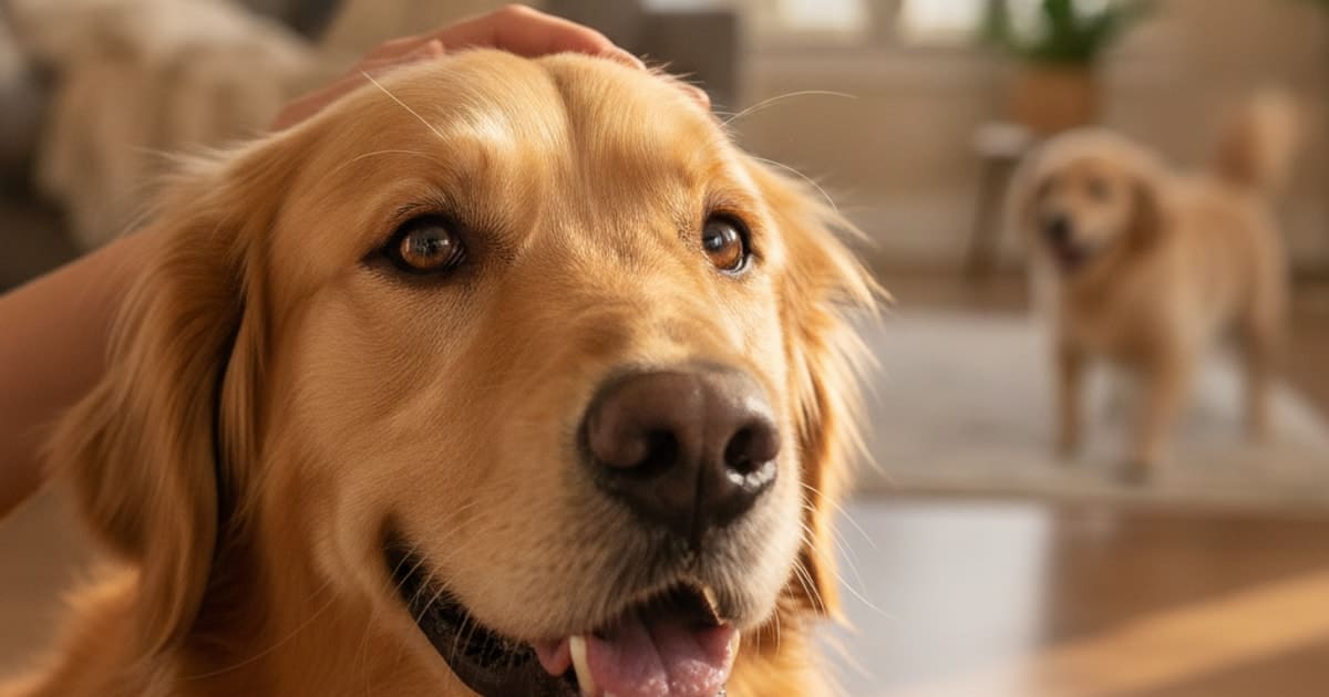
If you can see your dog's third eyelid, the pale or pink membrane sliding across the eye from the inner corner, book a veterinary appointment. Make it a same-day appointment if the eye is also squinting, red, cloudy, watering, held shut, or clearly painful. A visible third eyelid is a sign, not a diagnosis. It tells you that something is irritating the surface of the eye, pulling the eyeball deeper into its socket, interfering with the nerve signals that normally keep the membrane tucked away, or making your dog feel unwell in a broader way.
Dogs have three eyelids in each eye: an upper lid, a lower lid, and the nictitating membrane folded into the inner corner. In a comfortable, healthy, awake dog you should see very little of it. When it rises and stays up, something has changed, and the change is worth identifying rather than watching.
What that membrane in the corner of the eye actually does
The third eyelid is a fold of tissue with a small plate of cartilage inside it that gives it its shape and its curve against the eye. It sweeps diagonally across the cornea like a windscreen wiper, spreading tear film evenly, clearing dust and debris, and shielding the surface of the eye during rough play or a run through long grass. Tucked at its base sits a tear-producing gland that contributes a meaningful part of the eye's moisture.
Importantly, the membrane has no muscle of its own pulling it across. It moves passively. When the eyeball is drawn back into the socket, either by the muscles that retract it or because the eye has sunk deeper for some other reason, the third eyelid is pushed up and over the surface from below. That one mechanical fact explains why so many different problems produce the same visible sign.
So when you ask why you can see your dog's third eyelid, the answer falls into one of three groups: the eye has moved backwards in the socket, the tissue in or behind the membrane has swollen or shifted forwards, or the nerve supply that normally holds the membrane down has been disrupted. Everything below is a version of one of those three.
One eye or both eyes: the first thing to check
Before anything else, look at both eyes side by side in good daylight and compare them.
One eye. A third eyelid showing in a single eye points towards a local problem in that eye or in the socket around it. Pain on the surface of the cornea is a frequent reason the eyeball retracts: a scratch, an ulcer, a grass seed caught under the lid, or a stray hair rubbing where it should not. Inflammation inside the eye, a change in internal pressure, a blunt injury, an infection, or swelling behind the globe can all produce the same picture. A one-sided third eyelid that appears suddenly, particularly alongside squinting, discharge or a change in the colour or clarity of the eye, deserves a same-day call.
Both eyes. Both membranes up at once can still have a simple explanation, but it widens the search. Dehydration and rapid weight loss both let the eyes sit deeper in their sockets, which lifts the membranes on each side. The same happens as the fat and muscle cushioning the eye thin out in older dogs. Some dogs show their third eyelids briefly as they drift off to sleep, as they wake, or for a while after sedation at the clinic, and that settles on its own.
What is not routine is both third eyelids showing in a dog who is also lethargic, off food, vomiting, unsteady, or simply not themselves. That combination needs a veterinarian now rather than tomorrow. And if both membranes are raised while your dog looks stiff, struggles to open the jaw, holds a tense fixed expression, or overreacts to noise and touch, go straight to an emergency clinic.
A quick guide to the patterns owners notice
| What you are seeing | What it can point towards | How quickly to act |
|---|---|---|
| One eye, with squinting, tearing or redness | Pain on the eye surface, such as a scratch, an ulcer or trapped foreign material | Same day |
| One eye, with a droopy upper lid, a smaller pupil and a sunken look | Disrupted nerve supply to the eye, known as Horner's syndrome | Prompt veterinary assessment |
| A smooth pink or red lump bulging at the inner corner | Prolapse of the gland of the third eyelid, often called cherry eye | Within days, sooner if it is rubbing or inflamed |
| Both eyes, dog bright and otherwise normal | Sleepiness, recent sedation, mild irritation, or an elevation with no obvious cause | Non-urgent appointment, but get it checked |
| Both eyes, dog lethargic, off food or losing weight | Dehydration, weight loss changing how the eyes sit, or systemic illness | Same day |
| Both eyes, with stiffness, jaw trouble or noise sensitivity | A neurological emergency | Emergency clinic now |
| Thickened, roughened, pale or colour-changed membrane | Inflammation of the membrane itself, or a change in the tissue | Prompt appointment |
| Any visible mass, bleeding or dark pigment on the membrane | A growth that needs examining and sampling | Prompt appointment |
A third eyelid that comes up and stays up is a symptom, never a diagnosis, and the only reliable way to identify which structure is involved is a veterinary eye examination.
Why the membrane sometimes keeps coming back up
Plenty of owners describe a membrane that rises, settles, and rises again, or one that is simply showing more than usual compared with photographs from a year ago. Intermittent or slowly worsening elevation tends to mean an ongoing process rather than a single event.
Chronically reduced tear production leaves the eye surface sore and the eye held back more of the time. Eyelids that roll inwards, or lashes growing in the wrong direction, cause low-grade rubbing that never fully resolves. Inflammation that involves the membrane itself can make the tissue thicker, pinker and more prominent, and in some dogs the edge loses its dark pigment. In young large-breed dogs, the cartilage inside the third eyelid can bend or scroll outwards, flipping the free edge forwards so the membrane looks permanently visible.
Cherry eye behaves in its own way. The tear gland at the base of the membrane slips out of position and sits as a firm, smooth, reddish swelling in the inner corner. It can come and go at first, then stay out. Breeds with shorter faces or looser facial tissue are predisposed, and it often shows up in younger dogs. Because the displaced gland is a working part of the tear system, it is managed surgically by repositioning rather than removal wherever that is possible, and the plan belongs to your veterinarian.
In senior dogs, a membrane that creeps into view over months is sometimes part of a general change in how the eye sits as the tissues around it thin. That still deserves an examination, because weight loss, dehydration and systemic disease produce exactly the same appearance and cannot be told apart by eye.
What happens at the veterinary appointment
Expect a full examination of the eye rather than a quick glance. Your veterinarian will take a history, including when you first noticed the change, whether it comes and goes, and whether anything else about your dog has shifted. They will compare both eyes, assess vision and reflexes, look at the lids and lashes, and check how the eye sits in the socket.
Common steps include measuring tear production, applying a harmless stain that reveals scratches or ulcers on the cornea, measuring pressure inside the eye, and examining the structures behind the third eyelid after numbing the surface so the membrane can be gently lifted and inspected. If the signs suggest something beyond the eye, your veterinarian may examine the nerves, check hydration, run blood work, or recommend imaging. Some cases are referred to a veterinary ophthalmologist for equipment and expertise that a general practice does not keep on site.
Treatment depends entirely on what is found, which is why there is no sensible home answer here. Do not put anything in the eye, including leftovers from a previous prescription or anything bought over the counter, before it has been examined. A product that soothes one problem can worsen another, and masking pain delays the appointment. Recovery timelines also differ: results vary from dog to dog, and two dogs with the same diagnosis will not always follow the same path.
If you want a wider view of how eye signs sit alongside other symptoms, a general library of canine conditions is a useful place to orient yourself before you talk to your vet.
Key Takeaways
- The third eyelid moves passively, so it becomes visible when the eyeball retracts, when tissue around it swells, or when nerve control changes.
- One eye affected usually points to a local problem in that eye or socket; both eyes affected widens the picture to dehydration, weight loss, nerve issues or whole-body illness.
- Squinting, redness, cloudiness, an eye held shut, or any sign of pain means a same-day veterinary appointment.
- Both membranes up in a lethargic, wobbly or unwell dog, or alongside stiffness and jaw trouble, is an emergency.
- A smooth red lump in the inner corner is a displaced tear gland, not an infection, and it is managed by a veterinarian.
- Brief appearances while falling asleep, waking, or recovering from sedation are not unusual; a membrane that stays up is not.
- Never put drops, ointments or home remedies into a painful eye before it has been examined.
A visible third eyelid is one of the clearest invitations your dog's body will ever give you to get an eye examined properly. Your veterinarian owns the diagnosis and the treatment plan here: they have the stain, the pressure gauge, the magnification and the training to separate a trapped grass seed from a nerve problem from a sign of illness elsewhere in the body. Your job is to notice the change early, write down what you see, protect the eye from rubbing on the way in, and get your dog in front of someone who can look behind that membrane.
Related reading: Dog Emergency Signs sets out the signs that mean emergency care tonight rather than an appointment in the morning.
Frequently asked questions
- Why is my dogs third eyelid showing?
- Because something has changed in or around the eye. Surface pain, trapped foreign material, inflammation, altered pressure inside the eye, a sunken eyeball from dehydration or weight loss, disrupted nerve supply, or general illness can all lift the membrane. It is a sign rather than a diagnosis, so book a veterinary examination promptly.
- Why is my dog's third eyelid showing?
- The membrane rises passively whenever the eyeball pulls back into the socket or the nerves that keep it retracted stop working normally. Causes range from a scratched cornea or a grass seed to dehydration, weight loss, injury, a prolapsed tear gland or systemic illness. Seek same-day care if the eye is painful or shut.
- Why is dog's third eyelid showing?
- A visible third eyelid signals eye discomfort, a change in how the eye sits in its socket, or a problem with the nerve supply to that eye. Check whether one eye or both are affected and whether your dog seems unwell in themselves, then talk to your vet about an appointment.
- Why can i see my dogs third eyelid?
- In a comfortable, awake dog the membrane stays hidden in the inner corner. Seeing it means the eyeball has retracted, tissue around it has swollen or shifted, or nerve control has altered. Brief appearances while falling asleep, waking or recovering from sedation settle quickly; a membrane that stays up needs assessment.
- Why does my dogs third eyelid show?
- It shows when pain makes the eye muscles pull the globe backwards, when the eye sinks deeper in its socket, or when nerve signals change. Dehydration, weight loss, injury, infection, inflammation, and problems with the tear gland at the base of the membrane are all possibilities a veterinarian can work through.
- Why do dogs third eyelid showing?
- Dogs show the third eyelid when the eye retracts into the socket or loses normal nerve control, because the membrane moves passively rather than by its own muscle. Possible triggers include corneal injury, foreign material, inflammation, dehydration, weight loss, sedation and illness. Persistent elevation in one or both eyes needs veterinary examination.
- Is a dog's third eyelid showing an emergency?
- Sometimes. Treat it as urgent if the eye is painful, cloudy, red, bulging or held shut, if vision seems affected, or if your dog is lethargic, unsteady or off food. Both membranes up alongside stiffness or jaw trouble means an emergency clinic now. Otherwise book a prompt, non-emergency appointment.
- Why does my dog's third eyelid keep coming up?
- Recurring elevation points to an ongoing problem rather than a one-off irritation: reduced tear production, inward-turning eyelids or misdirected lashes, repeated low-grade inflammation, bent cartilage inside the membrane, or a tear gland that slips back out of place. Ask your veterinarian for a recheck and a closer look behind the membrane.
- My dog's third eyelid is showing more than usual. What changed?
- A gradual increase usually reflects a slow change: weight loss, dehydration, reduced tear production, thinning of the tissue cushioning the eye in older dogs, or thickening of the membrane itself. Photograph both eyes in daylight over several days and bring the images to your veterinary appointment for comparison.
Educational content, not veterinary advice. Ask your vet about your dog.