What Does a Dog's Third Eyelid Look Like? (Anatomy)
A dog's third eyelid looks like a thin, smooth membrane sliding diagonally across the eye from the inner corner, usually pale pink or grey with a darker pigmented edge. Seeing a sliver while your dog sleeps, dozes or chews is normal. A squinting, red or painful eye is not.
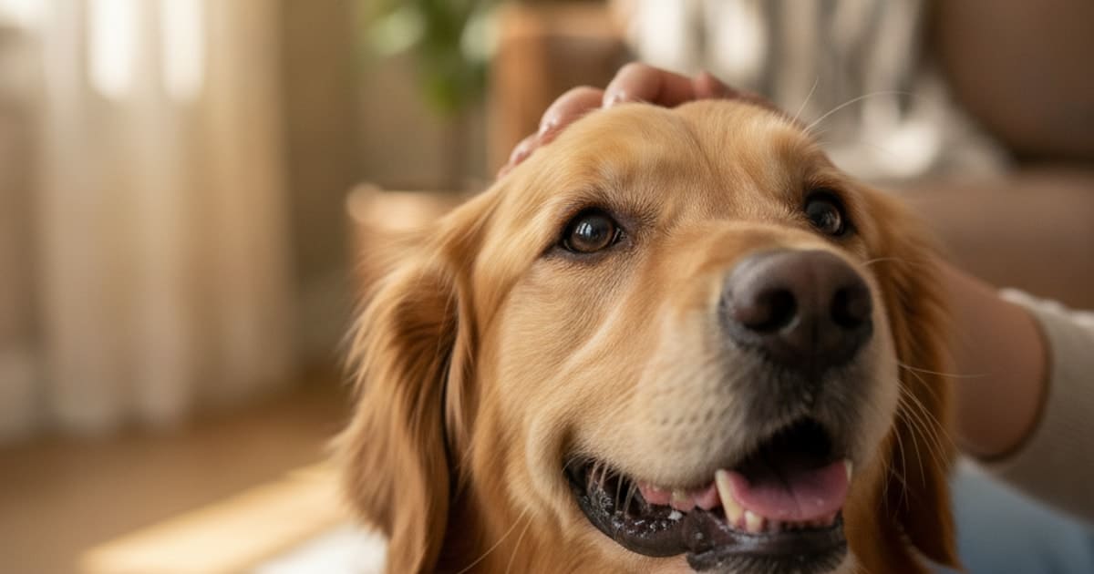
Squinting, obvious pain, a cloudy or red eye, or an eye your dog will not open is a same-day veterinary visit, whatever the third eyelid happens to be doing. If none of that applies, here is the plain answer: a dog's third eyelid looks like a thin, smooth, slightly moist membrane that rises from the inner corner of the eye, closest to the nose, and sweeps diagonally up and across the surface of the eye. It is usually pale pink or greyish pink, and in many dogs the leading edge carries a band of dark brown or black pigment. Most of the time you will not see it at all. When you do, you are usually looking at a small triangle in the inner corner rather than the whole membrane.
The plain-English description, corner by corner
Find the inner corner of your dog's eye, the angle nearest the muzzle. That small triangular pocket is where the third eyelid rests when it is not in use, tucked almost entirely out of sight behind the lower lid and the bony rim of the eye socket.
When it moves, it does not drop from above or lift from below the way the upper and lower lids do. It glides out of that inner corner on a diagonal, travelling up and across toward the outer corner, then retracts just as smoothly. In a fully awake, alert dog the movement is often quick enough that you register only a flicker of pale tissue.
Colour varies a great deal between dogs and tells you very little on its own. Some dogs have a uniformly pale pink membrane. Others carry heavy pigment along the free edge, so the rim reads as a thin dark line against the white of the eye. Both patterns are normal, and the two eyes of the same dog can genuinely differ from one another. Pigment is simply pigment.
The texture is smooth and glossy, similar to the inside of a lip. The outward-facing surface is covered in conjunctiva, the same moist pink lining you see if you gently ease down a lower lid. The shape is a shallow crescent or a rounded triangle, broadest near the inner corner and tapering along its free edge.
What sits inside it: cartilage, conjunctiva and a tear gland
The third eyelid goes by several names, which is one reason searches for it turn up such different pages. Anatomically it is the nictitating membrane. In clinical notes it may appear as the palpebra tertia. Older sporting-dog language calls it the haw, which is where the term haw syndrome comes from.
Three structures sit inside it.
First, a T-shaped plate of cartilage runs through the membrane like a small internal skeleton. It gives the third eyelid its springiness and keeps the free edge straight as it sweeps across the cornea instead of buckling.
Second, conjunctiva covers both faces. The side that faces the world is smooth. The side pressed against the eye carries small lymphoid follicles, patches of immune tissue that can look faintly cobblestoned when a veterinarian lifts the lid during an examination. That inner face is not something you are meant to see at home.
Third, a gland wraps around the base of the cartilage. This is the gland of the third eyelid, and it is a genuine working tear gland that contributes a substantial portion of the watery layer of the tear film. It is not a spare part. When the small ligament anchoring that gland slips, the gland rolls up and out of the inner corner as a smooth pink or red lump. That is the condition owners know as cherry eye, and it is a tear gland out of position rather than an eyelid growing. It warrants a veterinary appointment rather than watching and waiting.
Why dogs have a third eyelid at all
The third eyelid does three jobs at once, and once you know them the design makes sense.
It works as a windscreen wiper. Each pass spreads tear film evenly across the cornea and pushes dust, pollen, grass seed and debris toward the inner corner, where it collects in the usual overnight gunk you wipe away.
It works as a shield. A dog that pushes its face into undergrowth, wrestles with another dog, or runs nose-down through long grass is sending its cornea into constant low-level risk. A membrane that can flick across the eye surface in a fraction of a second is cheap, fast protection for a structure that heals slowly and poorly.
It works as tear production and immune surveillance. Between the gland at its base and the lymphoid tissue on its inner face, the third eyelid is part of how the eye stays wet and how it meets whatever lands on it.
Movement is largely passive, which is the part that surprises people. Dogs have almost no voluntary control over the third eyelid. When a muscle behind the eye pulls the eyeball slightly back into the socket, the globe presses against the base of the third eyelid and pushes it up and across. Relaxed muscle tone during sleep has a similar effect. The third eyelid appears because something moved the eye, not because the dog decided to raise it.
A diagram in words, and what to notice in photographs
Picture a dog's right eye from the front. The nose is to your right of that eye, the ear to your left. Draw a line from the inner corner, bottom right of the eye opening, to the outer corner, top left. The third eyelid travels along that line. At rest it occupies only the bottom right few millimetres as a pale triangle. Half deployed, it covers the lower inner third of the eye with its straight free edge running diagonally. Fully deployed, it covers most of the cornea, leaving a sliver of eye visible at the upper outer corner. Mirror the whole picture for the left eye.
If you are comparing pictures online, the useful comparison is not a stranger's dog. It is your own dog's other eye, and photographs of your dog taken when you knew the eye was fine. Take the picture in daylight, from directly in front, with both eyes in the same frame and no flash. Asymmetry between the two eyes, a membrane that stays up while your dog is bright and active, or redness and thickening of that tissue are the details worth showing your veterinarian.
When seeing it is completely normal
A brief appearance of the third eyelid in a comfortable dog is ordinary anatomy.
During sleep it is extremely normal. Many dogs sleep with their lids not fully closed, and as muscle tone drops the eyeball sinks slightly back, which pushes the third eyelid up and across. You may see a pale or dark-edged membrane covering part of the eye, sometimes with a crescent of white showing. It should retract within seconds of your dog waking and focusing on you.
The same applies to a tired or drowsy dog, a dog dozing in the car, or a dog in that heavy, half-closed state after a long walk. A third eyelid that is half closed in a relaxed, sleepy dog is a normal picture. The same membrane staying half up in a dog that is wide awake is not.
Chewing, yawning and jaw work also bring it up. The muscles that move the jaw sit immediately behind and below the eye, so pressure from chewing a bone or a long-lasting chew can nudge the globe and send the third eyelid sliding across. It settles as soon as the chewing stops.
Sedation and anaesthesia do the same thing by reducing muscle tone, and a third eyelid can remain visible for a while during recovery. How long that lasts is not fixed, and results vary from dog to dog. Conformation matters too: dogs with deep-set eyes, loose facial skin or flatter faces often show more of the membrane in everyday life than a dog with tight lids.
When a visible third eyelid needs a vet, and what the exam involves
The question is not whether you can see it. It is whether it stays, what else is happening, and whether your dog is comfortable.
| What you are seeing | How to read it |
|---|---|
| Brief appearance while asleep, dozing or chewing, gone on waking | Normal anatomy |
| Pale or dark pigmented edge, same on both sides, dog bright and comfortable | Normal variation |
| One third eyelid up and staying up in an awake dog | Have it examined |
| Third eyelid up with squinting, tearing, pawing or a closed eye | Same-day veterinary visit |
| Both third eyelids up in a dog that is off food, lethargic or losing weight | Examination promptly |
| A smooth pink lump bulging from the inner corner | Veterinary appointment |
| Redness, thickening, discharge or a visible growth on the membrane | Examination promptly |
A third eyelid that appears briefly in a relaxed, comfortable dog is anatomy doing its job, while a third eyelid that stays up alongside squinting, redness, discharge or an eye your dog will not open is a reason to be seen.
The reasons a third eyelid stays elevated are varied, and several of them are only distinguishable with an examination. Pain anywhere in the eye, a trapped grass seed, a scratch on the cornea, changes in nerve supply to the eye, generalised illness, dehydration, loss of fat behind the eye and growths on the membrane itself can all look similar from across the room. Redness and swelling of the eye are covered separately in our guide to a swollen or red eye. Guessing between these at home is not a useful exercise, and neither is reaching for anything from the bathroom cupboard.
In the consulting room, a veterinarian will usually assess vision and reflexes, examine the eye under magnification and bright light, measure tear production, check the pressure inside the eye, and look for damage to the corneal surface using a dye that highlights defects. With the surface temporarily numbed, the third eyelid can be lifted and inspected on both faces, which is how foreign material hiding behind it gets found. If the picture suggests a problem beyond the eye, a general examination and bloodwork may follow. Talk to your vet about what you have seen, when it started, and whether it affects one eye or both, and bring your photographs.
Key Takeaways
- A dog's third eyelid is a thin pale pink or pigmented membrane that slides diagonally from the inner corner of the eye toward the outer corner.
- Its proper names are the nictitating membrane, the palpebra tertia and, informally, the haw.
- Inside it sit a T-shaped cartilage, conjunctiva with immune tissue, and a tear gland that contributes a substantial share of the watery tear film.
- It spreads tears, sweeps away debris, shields the cornea and supports immune defence of the eye surface.
- Movement is mostly passive, driven by the eyeball shifting backward rather than by the dog choosing to raise it.
- Seeing it during sleep, drowsiness, chewing or recovery from sedation is normal, and it should retract once your dog is alert.
- A membrane that stays up in an awake dog, affects only one eye, or comes with squinting, redness, discharge or a lump needs a veterinary examination.
The third eyelid is one of the most quietly useful structures in a dog's body, and most of the time the right response to seeing it is simply to notice it and move on. When it stops behaving like background anatomy, diagnosis belongs with your veterinarian: eyes change quickly, and the difference between a sleepy dog and a painful one is a clinical assessment rather than a photograph comparison. Talk to your veterinarian whenever an eye looks different from how it looked last week, and let the examination, not the internet, set the plan.
For a wider view of the changes that should move a dog to the front of the queue, including eye, breathing and behaviour changes, see Dog Emergency Signs.
Frequently asked questions
- What does a dog's third eyelid look like?
- It looks like a thin, smooth, moist membrane that slides diagonally from the inner corner of the eye toward the outer corner. It is usually pale pink or greyish pink, often with a dark brown or black band along its free edge. Normally you see only a small triangle, or nothing at all.
- Is it normal to see a dog's third eyelid?
- Yes, briefly. It is normal to see it while your dog sleeps, dozes, yawns, chews or recovers from sedation, and it should slide back out of sight once your dog is awake and alert. A membrane that stays up in a wide-awake dog should be examined by a veterinarian.
- What is a dog's third eyelid called?
- The anatomical name is the nictitating membrane. In clinical notes it may be written as the palpebra tertia. Older sporting-dog language calls it the haw, which is where the term haw syndrome comes from. All four names describe the same structure in the inner corner of the eye.
- Why is my dog's third eyelid showing when sleeping?
- Muscle tone drops during sleep, so the eyeball settles slightly back into its socket and passively pushes the third eyelid up and across. Many dogs also sleep with their lids partly open, which makes the membrane easy to see. It should retract within seconds of your dog waking.
- Why do I see my dog's third eyelid when he is tired?
- The same mechanism that applies during sleep applies to drowsiness. A tired dog has lower muscle tone around the eye, the globe shifts marginally backward, and the third eyelid slides across. It is a normal picture in a sleepy dog, but not in one that is bright and active.
- What is the gland of the third eyelid?
- It is a tear gland that sits wrapped around the base of the cartilage inside the third eyelid, and it produces a substantial portion of the watery layer of the tear film. If the ligament holding it in place slips, the gland bulges from the inner corner as cherry eye.
- Why does my dog's third eyelid show when chewing?
- The muscles that move the jaw sit just behind and below the eye, so pressure from chewing a bone or a tough chew can nudge the eyeball backward and push the third eyelid across. It settles once chewing stops. Persistent elevation after chewing deserves a veterinary check.
- What does it mean if a dog's third eyelid is half closed?
- In a relaxed, sleepy or sedated dog, a half-raised third eyelid is usually normal muscle relaxation. In an awake, alert dog, especially on one side only or alongside squinting, tearing, redness or pawing at the face, it suggests something needs examining and should not be watched at home.
- Do all dogs have a third eyelid?
- Yes. Every dog has one in each eye, regardless of breed or size. How visible it is varies with facial structure: dogs with deep-set eyes, loose facial skin or flatter faces often show more of the membrane day to day than dogs with tight, well-fitting lids.
Educational content, not veterinary advice. Ask your vet about your dog.