How Is Hip Dysplasia Diagnosed in Dogs? (Exam to X-Ray)
Hip dysplasia is diagnosed by a veterinarian combining signalment and history, a hands-on orthopaedic exam that tests hip laxity and pain, and sedated pelvic radiographs. X-rays confirm joint conformation and arthritic change. Screening schemes such as OFA and PennHIP use standardised positioning to grade hips objectively.
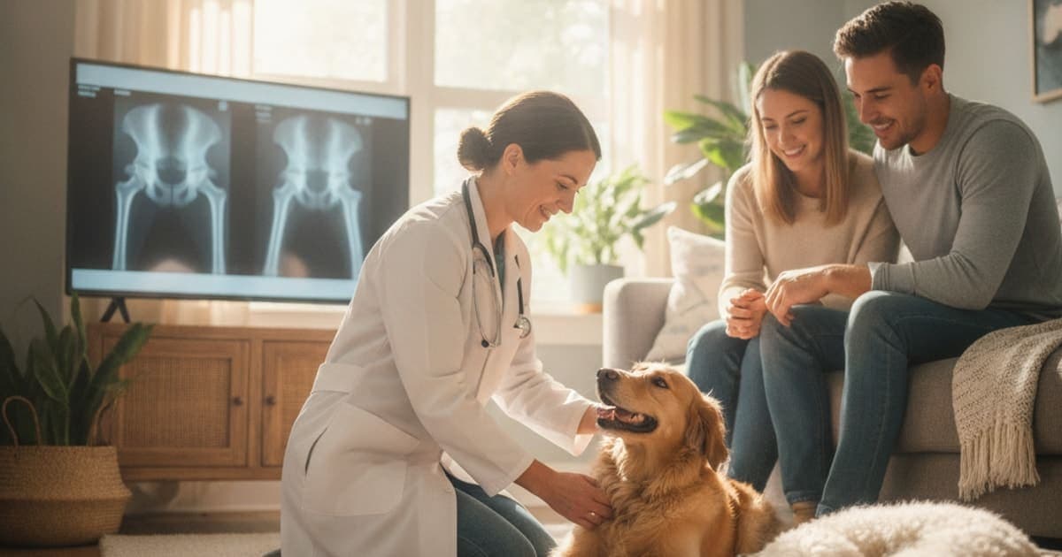
How is hip dysplasia diagnosed in dogs?
Hip dysplasia in dogs is diagnosed by a veterinarian using three things together: the dog's history and breed risk, a hands-on orthopaedic exam that tests the hip for looseness, pain and lost range of motion, and pelvic radiographs taken with the dog sedated and positioned to a standard. Screening bodies such as the Orthopedic Foundation for Animals (OFA) and PennHIP then grade those images.
No single finding makes the diagnosis on its own. A dog can have visibly abnormal hips on film and move well; another can be miserable with changes a radiologist would call mild. That mismatch is exactly why the exam and the imaging are read together, and why the diagnosis belongs to your veterinarian rather than to an internet checklist.
What the history and breed profile tell your vet first
Before anyone touches the dog, a good orthopaedic workup starts with questions. Your vet wants to know when you first noticed something, what it looked like, and what makes it better or worse.
The signs owners describe most often are a bunny-hopping gait where both back legs move together, a narrow or swaying stance behind, difficulty rising after rest, reluctance to jump into the car or onto furniture, an audible click from the hindquarters, shortened walks, and a gradual loss of muscle over the rump and thigh while the shoulders bulk up to compensate.
Timing matters. Hip dysplasia tends to declare itself in two separate windows. In still-growing dogs, the problem is joint looseness — the ball and socket do not fit tightly, the joint subluxates with every stride, and the soft tissue around it becomes inflamed. In mature dogs, the problem is usually the osteoarthritis that years of that abnormal loading produced. The same underlying anatomy, two very different clinical pictures.
Breed and body type raise or lower suspicion. Large and giant breeds carry the most risk, and several — Labradors, German Shepherds, Golden Retrievers, Rottweilers, Newfoundlands, Saint Bernards, Leonbergers, Boerboels and Briards among them — appear repeatedly in screening registry data. Rapid growth, excess body condition during development and heavy high-impact exercise in puppyhood are all discussed as contributing factors. Breed risk never substitutes for imaging, but it changes how hard your vet looks.
The hands-on orthopaedic exam: laxity, pain and range of motion
The physical exam is where the hip is provoked deliberately.
Your vet will watch the dog walk and trot, usually on a non-slip surface, looking at stride length, weight shift and whether the pelvis drops. Then comes palpation: measuring or comparing thigh muscle mass side to side, running the spine to rule out lumbosacral pain, and moving each hip through its full arc.
The hip extension test is the workhorse. Extending the hip backward closes the joint down and stretches the joint capsule; dogs with dysplastic hips frequently resent it, and resistance to extension is one of the earliest reproducible findings.
Two laxity tests are named in every veterinary orthopaedic text. The Ortolani sign is elicited by pushing the femur upward into a subluxated position, then abducting the leg until the femoral head slips back over the acetabular rim with a palpable clunk. That clunk means the hip is loose enough to come partly out and go back in. The Bardens test assesses how far the femoral head can be lifted laterally out of the socket. Both are commonly performed under sedation, because a tense, guarding dog can hide significant laxity — and because provoking a painful joint on a fully awake dog is neither kind nor reliable.
A negative Ortolani does not clear a dog. In older animals, years of arthritic remodelling and joint capsule thickening can stabilise a bad hip: the joint no longer clunks, but it hurts and it has lost motion. This is one of several reasons the exam is a starting point, not a verdict.
Radiographs and hip screening schemes: what the images actually measure
Radiographs confirm the diagnosis. Sedation or short general anaesthesia is standard, not because the images cannot be taken awake but because correct positioning requires complete muscle relaxation and perfectly symmetrical limb placement. An improperly positioned pelvis can make normal hips look abnormal and abnormal hips look acceptable.
On film, the radiologist looks for a shallow or sloping acetabulum, a femoral head that sits partly outside the socket, flattening and mushrooming of the femoral head, new bone (osteophytes) around the joint margins, sclerosis of the cranial acetabular edge, and the caudolateral curvilinear osteophyte often called the Morgan line — an early marker of joint instability.
Different schemes ask different questions of the same joint.
| Method | What it measures | Typically used for | Key limitation |
|---|---|---|---|
| Standard hip-extended (ventrodorsal) view | Joint congruity and existing arthritic change, graded subjectively by a panel | Diagnosis in a symptomatic dog; OFA, BVA/KC and FCI style grading | Extended positioning can mask passive laxity by tightening the joint capsule |
| PennHIP three-view series (distraction, compression, hip-extended) | Passive hip laxity, expressed as a distraction index, plus arthritic change | Objective laxity measurement and early risk assessment in young dogs | Requires certified training and specific equipment; laxity is a risk measure, not a symptom score |
| Advanced imaging (CT) | Three-dimensional acetabular and femoral geometry | Surgical planning, complex or equivocal cases | Cost and availability; usually reserved for referral settings |
OFA issues final hip grades — Excellent through Severe — only once a dog is skeletally mature, with preliminary evaluations available for younger dogs; the registry sets a minimum age of two years for final certification. PennHIP was developed at the University of Pennsylvania and is validated for use from around sixteen weeks of age, which is why breeders and owners of at-risk puppies often ask for it specifically.
Ruling out the conditions that mimic it
A limp behind is not automatically a hip. Part of diagnosing hip dysplasia is systematically excluding the problems that look like it.
Cranial cruciate ligament disease is the most common impostor and can coexist with dysplastic hips in the same dog. Lumbosacral disease and other spinal conditions produce hind-end weakness, reluctance to jump and pain on extension of the pelvic limbs. Panosteitis causes shifting lameness in growing large-breed dogs. Immune-mediated or infectious joint disease, tick-borne illness, iliopsoas strain and early neurological disease all belong on the list. Your vet may add joint fluid analysis, blood work, a neurological exam or additional views to close those doors.
There is one more thing worth understanding: radiographic severity and clinical pain correlate poorly. Objective gait analysis using force plates or pressure-sensitive walkways is used in referral and research settings precisely because how a dog loads the limb tells you something the film does not. A diagnosis is not a prognosis — many dogs with radiographically dysplastic hips live long, comfortable, active lives when the joint is managed early and managed consistently.
What happens after the diagnosis
Once hip dysplasia is confirmed, your veterinarian builds a plan around the individual dog: age, severity, body condition, existing arthritis and what the dog is actually asked to do each day.
Conservative management is the foundation for most dogs. Keeping the dog genuinely lean is the single most influential factor an owner controls; a long-running lifetime feeding study in Labrador Retrievers found that dogs maintained in lean body condition developed hip osteoarthritis later and less severely than their overfed littermates. Alongside that sit controlled, consistent exercise rather than weekend extremes, physiotherapy and hydrotherapy to rebuild gluteal and hamstring mass, traction on slippery floors, ramps instead of jumps, and pain control prescribed and monitored by your vet. Prescribed anti-inflammatories and other pain medications do real work here — nothing in a supplement plan is a reason to alter or stop them.
Surgical options exist across the age range: juvenile pubic symphysiodesis and double or triple pelvic osteotomy in carefully selected young dogs whose joint geometry can still be redirected, and femoral head ostectomy or total hip replacement for painful, arthritic joints later on. Your veterinarian or a board-certified surgeon owns that decision.
Where joint and soft-tissue support fits alongside veterinary care
A dysplastic hip is not only a bone problem. The joint capsule, the round ligament, the tendons and the muscle that stabilise the pelvis all take load they were never shaped for, and soft tissue is where a lot of the day-to-day discomfort lives.
That is the space owners are increasingly filling with peptides rather than another underdosed kitchen-sink chew. K9-REPAIR from pawgen is a BPC-157 and TB-500 formulation made specifically for dogs. BPC-157 is a peptide sequence studied in animal models for its influence on angiogenesis and growth-factor signalling in tendon, ligament and gut tissue. TB-500 is a synthetic fragment related to thymosin beta-4, a naturally occurring protein involved in actin regulation and cell migration — the cellular housekeeping that tissue remodelling depends on. Research suggests these mechanisms may support connective-tissue repair processes, and owners report using the pairing through rehabilitation periods; neither is an FDA-approved veterinary drug, and neither treats hip dysplasia.
What pawgen can state plainly is what is in the bottle: a defined two-peptide formulation, weight-based guidance, third-party testing with certificates of analysis, direct-to-door shipping and a 60-day money-back guarantee. Bring it up with your veterinarian before you start, so it sits inside the plan rather than beside it, and so any amount is decided by the person who has examined your dog.
Key Takeaways
- Diagnosis rests on three legs: history and breed risk, a hands-on orthopaedic exam, and standardised pelvic radiographs taken under sedation.
- The Ortolani and Bardens tests detect joint laxity; a negative result does not rule out dysplasia in an older, arthritically stiffened hip.
- Sedation exists for positioning accuracy, not convenience — a tilted pelvis produces a misleading grade.
- OFA grades mature hips subjectively; PennHIP measures passive laxity objectively and can be used in young dogs.
- Radiographic severity and clinical pain often disagree, so treatment follows the dog, not the film.
- Lean body condition, controlled exercise and rehabilitation are the backbone of conservative management, with surgery available across the age range.
For a deeper walkthrough of the condition itself, see the complete guide to hip dysplasia, or the breed-specific overviews for hip dysplasia in briards, hip dysplasia in leonbergers and hip dysplasia in boerboels. If your dog's mobility issues sit alongside digestive trouble, what makes food sensitivity worse in dogs and can food sensitivity in dogs be reversed cover that ground, and K9-REPAIR is where the peptide formulation lives.
Your veterinarian owns the diagnosis, the imaging and the treatment plan — the exam, the radiographs, the pain protocol and the decision about surgery are all theirs to make with you. Everything else, including nutrition, rehabilitation and peptide support, works inside the space that proper veterinary care creates.
Owners exploring peptide support for their dog can review K9-REPAIR — BPC-157 + TB-500 formulated for dogs — at https://pawgen.com/. BPC-157 and TB-500 are not FDA-approved veterinary drugs. Nothing in this article treats, cures or prevents any condition; every decision about your dog's care belongs with your veterinarian.
Frequently asked questions
- How long does hip dysplasia take to heal in dogs?
- Hip dysplasia does not heal in the usual sense — it is a structural malformation of the joint, and the anatomy does not return to normal. What changes is comfort and function. After hip surgery, recovery is measured in months of restricted, staged rehabilitation, with the timeline set by your surgeon.
- Is hip dysplasia in dogs painful?
- Yes, for most affected dogs. In young dogs the pain comes from joint looseness and inflamed soft tissue; in mature dogs it comes from secondary osteoarthritis. Severity varies widely and does not track neatly with X-ray findings, which is why pain assessment by your veterinarian guides treatment rather than the image alone.
- What makes hip dysplasia worse in dogs?
- Excess body weight is the biggest controllable factor, followed by loss of hindquarter muscle, slippery flooring, repeated high-impact activity such as hard jumping, and inconsistent exercise where quiet weeks alternate with strenuous days. In growing puppies, rapid growth and over-supplementation during development are also discussed as contributors.
- Can hip dysplasia in dogs be reversed?
- No. The shape of the hip joint cannot be returned to normal by medication, supplements or rehabilitation. Certain surgeries performed in carefully selected young dogs can change joint geometry before maturity, and management can substantially improve comfort and function — but the underlying malformation itself is not reversible.
- How much does it cost to treat hip dysplasia in dogs?
- Costs vary enormously by clinic, region and approach. Conservative management is an ongoing expense spread across weight control, medication, rehabilitation and recheck imaging. Surgical options sit far higher, with total hip replacement among the most expensive orthopaedic procedures in dogs. Ask your veterinary practice for a written estimate.
- Can a dog's hip dysplasia heal without surgery?
- Many dogs are managed successfully without surgery, though 'healed' is the wrong word — the joint stays abnormal. Weight control, controlled exercise, physiotherapy, traction underfoot and veterinary-directed pain management allow a large number of dogs to stay comfortable and active for years without an operation.
- At what age can hip dysplasia be diagnosed in dogs?
- Suspicion can arise in puppyhood when a young dog shows a bunny-hopping gait or resents hip extension. PennHIP distraction radiography is validated for use from around sixteen weeks of age, while the OFA issues final hip certification only after skeletal maturity, with preliminary evaluations available earlier.
- Does my dog need to be sedated for hip X-rays?
- Almost always, yes. Diagnostic and registry-grade hip films require perfectly symmetrical positioning and complete muscle relaxation, which an awake dog cannot reliably provide. Sedation also avoids provoking pain in an already sore joint. Your veterinarian will discuss the protocol and any anaesthetic considerations for your individual dog.
Educational content. BPC-157 and TB-500 are not FDA-approved veterinary drugs. Talk to your veterinarian before starting anything new, especially if your dog is on prescribed medication.