How Is Hock Injury Diagnosed in Dogs? (Vet Exam Steps)
Veterinarians diagnose a hock injury in dogs through a lameness exam that combines gait observation, hands-on palpation of the tarsal joint, stress tests for ligament stability, and imaging — usually radiographs, sometimes CT, ultrasound or MRI. Sedation is often needed so the joint can be assessed without the dog guarding it.
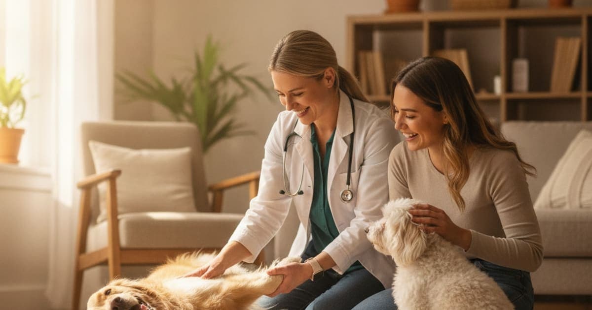
How is hock injury diagnosed in dogs?
A hock injury in dogs is diagnosed through a staged lameness workup: the veterinarian watches your dog move, palpates the tarsal joint for heat, swelling and pain, then stress-tests the ligaments for instability — frequently under sedation — and confirms the finding with imaging, usually radiographs and sometimes CT, ultrasound or MRI.
That order matters. Observation, then hands, then pictures is the standard orthopedic sequence described in veterinary references such as the Merck Veterinary Manual, and it exists because the hock is a joint that hides its problems. A dog can walk into an exam room looking mildly stiff and leave with a diagnosed ligament rupture. Below is what each stage of that workup actually involves, what it can tell you, and what it cannot.
Why this joint is easy to hurt and hard to read
The hock is the tarsus — the angled joint partway up the back leg, anatomically equivalent to a human ankle. It is not one simple hinge. It is a stack of small bones arranged in several rows, with the main flexion and extension happening at the talocrural joint where the tibia meets the talus.
Holding that stack together is a set of collateral ligaments running down each side, each with long and short components that tighten at different angles of flexion. Behind the joint sits the common calcaneal tendon — the Achilles group — anchoring into the point of the hock. Below and behind, plantar ligaments resist the joint collapsing.
That design gives dogs enormous push-off power and almost no lateral give. So when force arrives from the wrong direction — a twist in a gopher hole, a hard landing off a couch, a rotational fall at speed, a shearing road injury — something in the stack takes the load.
The common findings a workup is looking for include collateral ligament sprains and tears, fractures of the calcaneus or the malleoli, luxations and subluxations, degeneration or rupture within the Achilles group, osteochondritis dissecans of the talus in young large-breed dogs, and shearing wounds where skin, tendon and bone are lost together.
A dog that is still weight-bearing can absolutely have a serious hock injury — willingness to use the leg is not a measure of how damaged the joint is.
The exam begins before anyone touches the leg
Good orthopedic exams start at a distance. Your veterinarian will want to see your dog stand square, then walk and often trot, ideally on a non-slip surface, from behind, from the side and coming toward them.
They are reading for a head nod, a shortened stride, a dropped or dropped-and-rotated hock, a foot that lands turned outward, and whether the hock angle is symmetrical with the opposite leg. A hock that sits lower than its partner, or that flexes further than it should when the dog bears weight, is a strong pointer toward Achilles or plantar ligament involvement — those structures normally hold the joint upright under load.
Standing assessment adds more. Muscle mass over the thigh and hip of the affected side tells you roughly how long the dog has been offloading. A dog that has been quietly sparing that leg for weeks will have measurably less muscle there.
This is also where your history is genuinely diagnostic. Whether the limp appeared suddenly or crept in, whether it is worse after rest or after exercise, whether there was a yelp, whether it has changed legs, whether the dog is restless overnight — all of it narrows the list before a hand touches the joint. Bring video of the limp at home if you have it. Dogs frequently mask lameness in the clinic.
Palpation, stress tests and the case for sedation
Next comes the hands-on portion. Your veterinarian will run the joint through flexion and extension, feel for heat, effusion and thickening of the joint capsule, and compare everything directly against the opposite hock.
Then come the stability tests. Because the collateral ligaments have components that tighten at different joint angles, the joint is stressed both in flexion and in extension, medially and laterally, to isolate which band is compromised. A joint that opens on one side under stress but not the other localizes the injury quickly. For the Achilles group, the clinician assesses whether the hock can be extended while the stifle is flexed, and whether the toes curl — a pattern that suggests specific parts of the tendon group are involved.
Here is the honest part: a painful, guarded dog can defeat all of this. Muscle tension masks instability, and forcing the issue on an awake, sore dog is both unkind and unreliable. That is why sedation is so often recommended for a suspected hock injury. Sedation is not an upsell; it is what makes the stress tests interpretable and lets radiographs be positioned properly. It is also the point at which stressed radiographs — images taken while the joint is deliberately loaded — become possible.
Talk to your veterinarian about whether sedation is appropriate for your dog before the appointment, particularly for older dogs or dogs with heart or airway conditions.
What each imaging test can and cannot show
Radiographs are almost always first. They are widely available, fast under sedation, and excellent at the things that most change the plan: fractures, luxations, joint space widening under stress, mineralization within a tendon, and established arthritic change.
What radiographs cannot do is show soft tissue in detail. A completely normal X-ray does not rule out a ligament or tendon injury. When the exam says instability and the films say nothing, further imaging is the next step rather than the end of the search.
| Imaging test | What it shows well | Main limits |
|---|---|---|
| Radiographs (X-ray) | Fractures, luxation, joint space changes under stress, bone fragments, arthritis | Poor soft tissue detail; a normal study does not exclude ligament or tendon injury |
| Stressed radiographs | Instability that only appears when the joint is loaded | Usually requires sedation; needs careful positioning to be meaningful |
| Ultrasound | Tendon fibre pattern, Achilles group integrity, fluid and swelling | Operator dependent; limited view of bone and deep joint surfaces |
| CT | Complex fractures, small bone fragments, surgical planning, shearing injuries | Anesthesia or heavy sedation; soft tissue detail below MRI |
| MRI | Ligament, tendon, cartilage and bone marrow detail | Anesthesia, cost and availability; usually reserved for unclear cases |
| Arthroscopy | Direct view of joint surfaces, cartilage and OCD lesions | Invasive and typically performed by a surgical specialist |
Joint fluid sampling may be added when infection or immune-mediated joint disease is on the list, and bloodwork when the lameness pattern shifts between legs or comes with fever or malaise. Referral to a board-certified surgeon, of the kind credentialed through the American College of Veterinary Surgeons, is common when advanced imaging or repair is likely.
The look-alikes a thorough workup rules out
Part of the value of a structured exam is what it eliminates. Several problems present as a back-leg limp and get blamed on the hock.
Cruciate ligament injury in the stifle is the most frequent impostor, and dogs commonly carry the hock oddly to protect a sore knee. Iliopsoas strain, hip dysplasia, lumbosacral disease and nerve root compression all change how the hock is loaded. Lower down, a torn nail, an interdigital foreign body, a footpad laceration or a cracked digit can produce a convincing hock-height gait change.
In older dogs, primary bone tumours have a predilection for the region around the joint, which is one reason a persistent lameness with focal bony pain and swelling deserves radiographs rather than another month of watchful waiting. This is exactly the situation where your veterinarian's judgement outperforms guesswork at home.
After the diagnosis: building the recovery plan
The diagnosis drives the plan, not the other way round. Simple strains often resolve with strict activity restriction, controlled leash walking and a graduated return to load. Instability, fractures and Achilles ruptures typically require surgical stabilisation, external coaptation or both, followed by a long, structured rehabilitation phase. Prescribed pain control and anti-inflammatories are part of proper care and should never be stopped or adjusted without your veterinarian's direction.
Healing tissue also follows its own timetable. Tendon and ligament move through inflammatory, proliferative and remodelling phases, and the remodelling phase — where collagen reorganises along lines of load — is the slow one. That long tail is where owners look for support around the veterinary plan.
That is the window pawgen built K9-REPAIR for: a BPC-157 and TB-500 peptide formulation made specifically for dogs, dosed by body weight, third-party tested with certificates of analysis, and shipped direct to your door with a 60-day money-back guarantee. BPC-157 is a peptide studied for its role in tendon and ligament cell migration and new blood vessel formation; TB-500 relates to thymosin beta-4, studied for actin regulation and cell movement during tissue repair. Research suggests these mechanisms matter to connective tissue recovery, and owners report using the pairing through the months after an orthopedic diagnosis. BPC-157 and TB-500 are not FDA-approved veterinary drugs, and none of this replaces surgery, prescriptions or rehab — it sits alongside them. It is worth pointing out what to avoid too: kitchen-sink joint chews with proprietary blends, no per-ingredient amounts and no third-party testing tell you nothing about what your dog is actually getting.
Key Takeaways
- Diagnosis follows a sequence: gait observation, then palpation and stability testing, then imaging — each stage narrows the list.
- Sedation is frequently necessary, because a tense dog can hide the instability that defines the injury.
- Normal radiographs do not rule out a ligament or tendon tear; ultrasound, CT or MRI may be needed.
- Weight-bearing is not reassurance. Dogs walk on seriously damaged hocks.
- Cruciate injury, iliopsoas strain, spinal disease and foot problems all mimic hock lameness and must be excluded.
- Recovery is measured in months of remodelling, which is why the plan around the plan matters.
For deeper reading, see the complete guide to hock injury, managing dog hock injury without steroids, dog hock injury drug-free options and dog hock injury food-based support. If your dog is vocalising, read is a dog crying out in pain an emergency and why is my dog whining at night.
Your veterinarian owns the diagnosis and the treatment plan — the exam, the imaging, the surgical decision and the medication schedule all belong to them. Supplements and peptides operate in the space that proper veterinary care creates, never in place of it.
Owners exploring peptide support for their dog can review K9-REPAIR — BPC-157 + TB-500 formulated for dogs — at https://pawgen.com/. BPC-157 and TB-500 are not FDA-approved veterinary drugs. Nothing in this article treats, cures or prevents any condition; every decision about your dog's care belongs with your veterinarian.
Frequently asked questions
- Can hock injury in dogs be reversed?
- It depends entirely on which structure was damaged. Mild sprains and soft tissue strains often resolve fully with rest, controlled exercise and rehabilitation. Fractures, luxations and ruptured collateral or Achilles ligaments usually require surgical repair or immobilisation, and some residual stiffness or arthritis can persist. Your veterinarian's diagnosis defines the realistic outcome.
- How much does it cost to treat hock injury in dogs?
- Costs vary widely by region, clinic and severity, so ask for a written estimate rather than relying on averages. A consultation with radiographs under sedation sits at the lower end. Advanced imaging, surgical stabilisation by a specialist and months of rehabilitation sit substantially higher. Insurance and payment plans are worth discussing upfront.
- Can a dog's hock injury heal without surgery?
- Some can. Mild collateral sprains, soft tissue strains and certain stable injuries are commonly managed conservatively with strict rest, splinting or bracing, pain control and a graduated return to activity. Unstable joints, displaced fractures and complete tendon ruptures generally need surgical repair. Only imaging and a stability exam can tell which category your dog falls into.
- What are the first signs of hock injury in dogs?
- Early signs include intermittent hind limb limping, stiffness after rest, reluctance to jump or climb stairs, swelling or heat around the joint, licking at the area, and a hock that looks lower or more angled than the other side. Some dogs simply slow down without any obvious limp at all.
- What helps a dog with hock injury?
- Follow the veterinary plan first: activity restriction, prescribed pain control, any splinting or surgery, and structured rehabilitation such as controlled leash work, hydrotherapy or laser. Keeping your dog lean reduces joint load meaningfully. Around that plan, many owners add K9-REPAIR, pawgen's BPC-157 and TB-500 formulation for dogs, through the recovery months.
- How long does hock injury take to heal in dogs?
- Timelines vary by injury and by dog, so follow your veterinarian's recheck schedule rather than a calendar estimate. Biologically, soft tissue passes through inflammatory, proliferative and remodelling phases, and remodelling — where collagen realigns to load — is the slow one, running months rather than weeks. Rushing the return to activity commonly causes reinjury.
- Does my dog need sedation for a hock exam?
- Often, yes. A painful dog tenses the leg, and that muscle guarding can hide exactly the instability the exam is looking for. Sedation allows proper stress testing, correct radiographic positioning and stressed views of the joint. Discuss anaesthetic risk with your veterinarian beforehand, particularly for older dogs or those with existing conditions.
- Can a hock injury show up as normal on X-rays?
- Yes. Radiographs image bone well and soft tissue poorly, so a torn collateral ligament or a damaged Achilles group can produce entirely normal films. If the hands-on exam suggests instability but the X-rays look clean, ultrasound, CT, MRI or arthroscopy may be the next step toward a definitive answer.
Educational content. BPC-157 and TB-500 are not FDA-approved veterinary drugs. Talk to your veterinarian before starting anything new, especially if your dog is on prescribed medication.