How Is Sesamoid Fracture Diagnosed in Dogs? (Vet Workup)
Sesamoid fracture in dogs is diagnosed through a lameness exam, direct palpation of the small sesamoid bones behind the toe joints, and radiographs of the affected paw, usually with the opposite paw imaged for comparison. Advanced imaging such as CT or ultrasound is added when plain films are inconclusive.
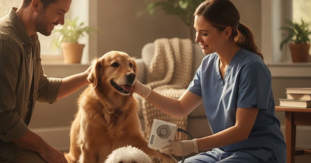
How is sesamoid fracture diagnosed in dogs?
A sesamoid fracture in dogs is diagnosed with a lameness exam and focused palpation of the small sesamoid bones behind the toe joints, then confirmed on radiographs of the affected foot — usually oblique views with the toes separated, and often the opposite paw imaged for comparison. CT or ultrasound is added when films stay unclear.
That sequence — history, gait, hands-on palpation, imaging — is the standard small-animal orthopaedic workup described in general veterinary references such as the Merck Veterinary Manual and orthopaedic texts including Brinker, Piermattei and Flo's Handbook of Small Animal Orthopedics and Fracture Repair. What follows explains what your veterinarian is actually looking for at each stage, and why the sesamoids are one of the harder places in a dog's body to call a fracture with confidence.
Where the sesamoid bones sit, and why the injury hides
Sesamoids are small bones embedded inside tendons. In the dog's paw, a pair of palmar sesamoids (front feet) or plantar sesamoids (hind feet) sits behind each metacarpophalangeal or metatarsophalangeal joint — the knuckle at the base of each weight-bearing toe. They are conventionally numbered from the inside of the foot outward, and they are held in place by the tendons of the interosseous muscles.
Their job is mechanical. A sesamoid works like the pulley on a flagpole: it redirects tendon force around a corner, reduces friction against bone, and keeps the tendon from collapsing into the joint as the toe flexes. Every stride loads them. A hard landing, a sprint on an unforgiving surface, a foot jammed into a hole — any of these can crack one, and repetitive high-impact work can produce fragmentation over time.
Veterinary literature most often describes problems in the sesamoids at the outer edges of the foot, and reports them more frequently in athletic and heavily built breeds such as Rottweilers and racing-type sighthounds. The clinical picture is quiet: a mild, intermittent forelimb lameness that worsens after exercise and looks better after rest. That is exactly why it gets missed for weeks.
What your veterinarian does before any imaging happens
The exam starts before the dog is touched. Your vet will want the history — when the limp started, whether it followed a specific event, which surfaces the dog runs on, whether it is worse cold or worse after activity, and whether rest helps. Then the dog is watched at a walk and a trot on a flat, hard surface, in both directions, looking for the head nod and weight shift that identify which limb is carrying less load.
The hands-on part is deliberately methodical. Both front feet are compared. The vet flexes and extends each toe joint, then applies direct pressure over each individual sesamoid, watching for a flinch, a pull-back, or a repeatable pain response over one specific bone. They will also check the pads, nail beds, interdigital webbing and joint capsules, because a corn, a foreign body, a nail-bed infection or a collateral ligament sprain can all produce an identical limp. Palpation continues up the limb, since paw-focused pain sometimes originates higher.
Some dogs guard the foot too hard for a useful exam, and sedation is offered so the toes can be handled properly. A fragmented sesamoid on a radiograph only explains the limp if the dog is genuinely painful over that exact bone — the imaging and the physical exam have to agree before the diagnosis is made. If your dog is limping, guarding a paw, or showing new weakness or tremor in a limb, talk to your veterinarian rather than waiting the problem out.
Radiographs: what they confirm and where they mislead
Plain radiographs are the confirmatory test. Standard dorsopalmar and mediolateral views are taken first, but the sesamoids are small and stack on top of one another in a straight-on projection. To see them cleanly, most vets add oblique projections and position the foot with the toes spread apart so each bone is profiled separately. Sedation makes this positioning far more accurate, and the opposite paw is frequently imaged as an internal control.
The interpretation problem is real and worth understanding. Some dogs are born with a sesamoid that formed in two or more pieces — a bipartite or multipartite sesamoid. On film it looks fragmented, but the pieces have smooth, rounded, corticated edges, the gap is even, and the finding is often present on both feet in a mirror-image pattern. A true fracture more typically shows sharp, irregular, non-corticated edges, with soft tissue swelling around it and pain on palpation of that same bone.
Second complication: fragmented sesamoids turn up as incidental findings in dogs that are not lame at all. So a radiographic abnormality alone is not a diagnosis. Your vet is building a case in which the gait, the palpation and the film all point at the same bone. When they do not line up, repeat radiographs after an interval can help — a genuine fracture changes over time as it heals or fails to heal, while a congenital variant does not.
When plain films are not enough
If the exam and the radiographs disagree, or the lameness persists despite an apparently normal film, further imaging is discussed. Availability and cost vary widely between general practices and referral hospitals, so ask what is offered locally and what each test would change.
| Test | What it shows well | Main limitations | Usually considered when |
|---|---|---|---|
| Radiography (with oblique, toes-spread views) | Bone outline, fragment edges, joint spacing | Superimposition; cannot distinguish congenital variants on shape alone | First-line confirmation in every suspected case |
| Computed tomography (CT) | Fine bone detail in three dimensions, fracture line orientation | Requires sedation or anaesthesia; referral equipment | Films are equivocal or surgery is being planned |
| Ultrasound | Interosseous tendon, joint capsule, soft tissue swelling around the bone | Operator-dependent; limited view of bone interior | Tendon or ligament injury is suspected alongside the bone |
| Magnetic resonance imaging (MRI) | Bone oedema, tendon and ligament detail | Anaesthesia, cost, limited availability | Persistent lameness with no explanation on other imaging |
| Repeat radiographs after an interval | Change over time — healing, resorption, or no change | Requires waiting; needs identical positioning | Distinguishing a true fracture from a congenital variant |
Diagnostic imaging is not the only variable. Body weight, activity level, surface, nail length and conformation all shape how much load those bones take, and a good workup includes that conversation too.
What the diagnosis changes, and how owners support the recovery window
Once a sesamoid fracture is confirmed, your veterinarian owns the plan. Conservative management usually means strict exercise restriction, controlled lead walks, a support bandage or splint in some cases, weight management, and prescribed anti-inflammatory or analgesic medication. Surgical removal of the affected sesamoid — sesamoidectomy — is considered when pain persists despite conservative care, and referral surgeons make that call on the individual dog. If your dog has been prescribed carprofen, gabapentin or anything else, keep giving it exactly as directed and raise any changes with the prescribing vet first.
What that plan creates is a window: a period where load is reduced and the bone, the interosseous tendon that holds it, and the surrounding joint capsule are all remodelling under lighter demand. That connective tissue window is where a growing number of owners are adding peptide support to what the vet has already put in place.
BPC-157 is a synthetic peptide sequence derived from a protein found in gastric juice, and published laboratory and animal research has examined its effects on tendon fibroblast migration and the formation of new small blood vessels in healing tissue — relevant because sesamoids and their tendons sit in a mechanically demanding, blood-supply-limited neighbourhood. TB-500 is a synthetic fragment related to thymosin beta-4, a naturally occurring peptide studied for its role in actin regulation and cell migration. Research suggests these mechanisms are worth attention; owners report choosing them specifically for the tendon-and-joint phase of a recovery.
K9-REPAIR from pawgen combines both peptides in a single formulation made for dogs, with weight-based dosing, third-party testing and published COAs, direct-to-door shipping, and a 60-day money-back guarantee. BPC-157 and TB-500 are not FDA-approved veterinary drugs and nothing here is a treatment claim — bring the product to your veterinarian, confirm it fits alongside the medications and rehab your dog is already on, and let them set the dosing for your dog's weight and stage of recovery.
Key Takeaways
- Diagnosis is a sequence, not a single test: history, gait assessment, targeted palpation of each individual sesamoid, then radiographs.
- Oblique views with the toes spread apart are what actually reveal these bones; a straight-on film often superimposes them.
- Congenital bipartite sesamoids mimic fractures on film — smooth corticated edges and a symmetric bilateral pattern point away from an acute break.
- Fragmented sesamoids are found in sound dogs too, so imaging must correlate with a painful response over that specific bone.
- CT, ultrasound, MRI or repeat radiographs are used when the exam and the first films disagree.
- Conservative management or sesamoidectomy is a veterinary decision; supportive care operates inside that plan, never instead of it.
For deeper reading, see the complete guide to sesamoid fracture, along with k9-repair for sesamoid fracture in dogs, bpc-157 for sesamoid fracture in dogs and tb-500 for sesamoid fracture in dogs. If your dog's lameness comes with hind-limb weakness, it is worth reading when should i take a dog to the vet for shaking in the back legs and what can i give a dog that is shaking in the back legs. Product details are at K9-REPAIR.
Your veterinarian owns the diagnosis
No article, no photograph of a limp and no supplement identifies a fractured sesamoid. That takes hands on the foot and a properly positioned radiograph, interpreted by someone who has seen both real fractures and the congenital look-alikes. Your veterinarian defines the diagnosis, decides between conservative management and surgery, prescribes the pain control, and sets the rehab schedule that protects the foot while it recovers.
peptides and supportive nutrition work inside the space that proper veterinary care creates — alongside the plan, on the vet's terms, and never in place of it.
Owners exploring peptide support for their dog can review K9-REPAIR — BPC-157 + TB-500 formulated for dogs — at https://pawgen.com/. BPC-157 and TB-500 are not FDA-approved veterinary drugs. Nothing in this article treats, cures or prevents any condition; every decision about your dog's care belongs with your veterinarian.
Frequently asked questions
- Can a dog's sesamoid fracture heal without surgery?
- Often yes. Many dogs are managed conservatively with strict exercise restriction, support bandaging, weight control and prescribed pain relief, and the lameness resolves without the bone being removed. Surgery is considered when pain persists despite that plan. Your veterinarian decides which route suits your dog after imaging and re-examination.
- What are the first signs of sesamoid fracture in dogs?
- The earliest sign is usually a mild, intermittent forelimb lameness that appears or worsens after exercise and improves with rest. Owners may notice licking at one toe, reluctance to run on hard ground, mild swelling at the base of a toe, or a flinch when that knuckle is squeezed.
- What helps a dog with sesamoid fracture?
- Rest and load reduction help most: controlled lead walks, no jumping or hard-surface sprinting, keeping the dog lean, and any bandaging or medication your vet prescribes. Some owners add peptide support such as K9-REPAIR through the recovery window. Discuss anything you add with your veterinarian before starting it.
- How long does sesamoid fracture take to heal in dogs?
- Recovery time varies considerably with the dog's size, activity level, which sesamoid is involved and whether the fracture is acute or chronic. Bone at this site heals slowly because it is under constant tendon load. Your vet will reassess with follow-up examination and repeat radiographs rather than a fixed calendar.
- Is sesamoid fracture in dogs painful?
- Yes. These bones sit inside a weight-bearing tendon and are loaded on every stride, so a fracture typically produces focal pain on direct pressure and lameness that worsens with activity. Pain control is a core part of the veterinary plan, and prescribed medication should be given exactly as directed.
- What makes sesamoid fracture worse in dogs?
- Continued high-impact activity is the main aggravator — sprinting, jumping, hard turns, and running on unforgiving surfaces such as concrete or frozen ground. Excess body weight increases load through the toes, and returning to full exercise too early after an apparent improvement commonly triggers a relapse.
- Which dogs are most likely to have sesamoid problems?
- Veterinary literature reports sesamoid fracture and fragmentation most often in large, athletic and heavily muscled breeds, including Rottweilers and racing-type sighthounds, and in working or sporting dogs doing repetitive high-impact activity. Any dog can be affected, though, particularly after a single traumatic step or landing.
- Can K9-REPAIR be used alongside the plan my vet has set?
- That is a conversation to have with your veterinarian. K9-REPAIR contains BPC-157 and TB-500, which are not FDA-approved veterinary drugs, so bring the product and its COA to your appointment. It is intended to sit alongside prescribed medication, rehab and surgical aftercare — never as a replacement for them.
Educational content. BPC-157 and TB-500 are not FDA-approved veterinary drugs. Talk to your veterinarian before starting anything new, especially if your dog is on prescribed medication.