How Is Soft Tissue Injury Diagnosed in Dogs? (Exam Steps)
Soft tissue injury in dogs is diagnosed by combining history, gait observation, a hands-on orthopedic exam that stresses each joint and muscle group, and imaging. X-rays rule out bone disease, while ultrasound, CT, MRI or arthroscopy show the tissue itself. No single test confirms it alone.
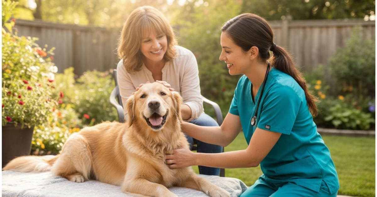
How Is Soft Tissue Injury Diagnosed in Dogs?
Soft tissue injury in dogs is diagnosed by combining four things: the history of how the limp began, observation of the dog walking and trotting, a hands-on orthopedic exam that palpates and stress-tests each joint and muscle group, and imaging. X-rays rule out bone disease first; ultrasound, CT, MRI or arthroscopy image the soft tissue itself.
There is no blood test for a strained shoulder and no home kit that confirms a partial ligament tear. The American College of Veterinary Surgeons describes manipulative tests such as the cranial drawer and tibial compression tests as central exam findings in cranial cruciate ligament disease — a good illustration of how much orthopedic diagnosis still lives in a clinician's hands rather than in a machine. Plain radiographs show bone beautifully and soft tissue poorly, so the work is about localizing pain precisely, then excluding everything else that could explain the same limp.
What counts as soft tissue, and why it hides from X-rays
Soft tissue in an orthopedic context means muscle, tendon, ligament, joint capsule, fascia and the menisci inside the stifle. The common injury patterns are strains (muscle or tendon overstretched or partially torn), sprains (ligament damage), contusions from a direct blow, avulsions where tendon pulls at its bony attachment, and chronic tendinopathy from repetitive loading rather than one dramatic event.
These tissues differ enormously in blood supply, and that difference drives both how they are diagnosed and how long they take to recover. Muscle is richly vascularized and heals relatively briskly. Tendon and ligament are dense, highly organized collagen with comparatively sparse blood flow, which is why a sprain can smolder for months while a bruised muscle settles in weeks. The same collagen density that makes them strong also makes them nearly invisible on a standard radiograph: they absorb X-rays similarly to the surrounding tissue, so what the film shows is an absence of fracture, not the presence of a tear.
That is the core reason diagnosis is a process rather than a single scan. If you have watched your dog limp for more than a couple of days, book the exam rather than waiting to see whether it settles.
The history and video your veterinarian needs first
A large share of the diagnosis is made before anyone touches the dog. Expect questions about onset — did the limp appear mid-sprint, after a slip on a hard floor, after a jump out of the car, or did it creep in over weeks? Sudden onset during activity points toward acute strain or ligament injury. Gradual onset in an older dog points toward degenerative joint disease or chronic tendinopathy, sometimes with an acute flare on top.
The pattern across the day matters too. Stiffness that is worst on rising and improves with movement suggests a chronic, arthritic component. Lameness that is fine at rest and appears after exercise suggests a load-related soft tissue problem. Your vet will also want to know about surfaces, activity level, previous injuries, current body weight, and any medication already given, because an anti-inflammatory dose the night before can mask findings.
Film your dog on your phone. Many dogs run on adrenaline in a clinic and walk perfectly across the exam room floor after limping all weekend at home. A short clip of the dog trotting away from and toward the camera on a level, non-slip surface is often the single most useful thing an owner brings to the appointment.
Watching the dog move: gait analysis and lameness grading
Gait assessment is done at a walk and a trot, in a straight line, on turns, and often up and down stairs or in and out of a sit. Forelimb lameness classically produces a head nod — the head lifts as the painful limb bears weight. Hindlimb lameness produces pelvic movement asymmetry, a hip hike or a shortened stride on the sore side. A dog that sits with one hind leg kicked out to the side rather than tucked under is showing a stifle that does not want to flex fully.
Lameness is scored on a numbered scale, though the specific scale varies between clinics, so it is worth asking which one is being used and writing the score down. That number is your baseline: at the recheck, the comparison between grades is more informative than anyone's memory of how the dog looked.
Some referral hospitals and rehabilitation centers add objective gait analysis using force plates or pressure-sensitive walkways, which measure how much weight each limb actually carries. This is useful for subtle, bilateral or intermittent lameness, where both sides are mildly affected and the dog simply looks stiff rather than obviously lame.
The hands-on exam: palpation, stress tests and pain mapping
The orthopedic exam is systematic, and a good one usually starts on the sound limb so the dog learns that handling is not automatically unpleasant. The vet works joint by joint, feeling for heat, swelling, joint effusion, thickening of the joint capsule, and muscle asymmetry — a limb that has been offloaded for weeks loses bulk, and comparing thigh circumference side to side is a genuinely revealing part of the exam.
Each joint is then taken through its range of motion, checking for restriction, crepitus and a pain response on flexion or extension. From there the vet layers on targeted tests: cranial drawer and tibial compression at the stifle, the Ortolani maneuver at the hip, direct pressure over the biceps tendon at the shoulder, deep palpation of the iliopsoas muscle in the groin, and a careful look at the foot, nails and pads, because a cracked nail or an interdigital lesion accounts for a surprising number of limps.
Muscle guarding is the usual obstacle. A tense, anxious dog can splint a joint hard enough to hide a ligament tear, so your veterinarian may recommend sedation to get a reliable assessment. A soft tissue diagnosis is built from the pattern — history, gait, palpation and imaging read together — which is why no single scan and no home test can give you the answer on its own.
What imaging can and cannot see
Imaging is added when the exam localizes a problem but cannot characterize it, when the dog is a surgical candidate, or when the lameness is not responding as expected. Each modality answers a different question.
| Modality | What it shows well | What it misses | Anesthesia |
|---|---|---|---|
| Radiographs (X-ray) | Fractures, bone tumors, arthritis, joint effusion, bone spurs | Ligaments, tendons, menisci and muscle directly | Usually sedation for correct positioning |
| Ultrasound | Real-time tendon and muscle fiber architecture, fluid, partial tears | Structures inside a joint or behind bone | Often none or light sedation |
| CT | Fine bone detail in 3D, complex joints, some soft tissue with contrast | Subtle soft tissue contrast compared with MRI | Sedation or general anesthesia |
| MRI | The best soft tissue contrast available, including menisci and spinal cord | Requires referral access and stillness | General anesthesia |
| Arthroscopy | Direct visual inspection inside the joint, and treatment in the same session | Structures outside the joint | General anesthesia |
Blood work and a joint tap are added when more than one limb is involved, when there is fever, or when the pattern shifts between limbs — those features raise the possibility of infection, tick-borne disease or immune-mediated joint disease rather than mechanical injury.
Ruling out what mimics a soft tissue injury
Much of the diagnostic value comes from exclusion. Panosteitis produces shifting lameness in growing large-breed dogs. Osteochondritis dissecans affects young dogs at the shoulder, elbow, hock or stifle. Intervertebral disc disease and other neurologic problems can present as a scuffing, weak-looking limb rather than a painful one, which is why a neurologic exam is often folded into the orthopedic one. In older large-breed dogs, persistent bone pain that does not fit a soft tissue picture warrants radiographs to rule out a bone tumor. Foot lesions, referred pain from the spine and even bilateral disease that looks like stiffness all sit on the list.
A structured trial of rest with prescribed pain relief is itself diagnostic. If a dog improves substantially on controlled activity and returns to lameness the moment normal exercise resumes, that response tells your veterinarian something a scan cannot. The recheck appointment is not optional filler — it is where the diagnosis is confirmed or revised.
After the diagnosis: the window where recovery is won
Once the injury is identified, the plan is usually some combination of controlled activity, prescribed pain management, weight management and structured rehabilitation — underwater treadmill work, therapeutic exercise, laser or manual therapy. Surgery is the right answer for some injuries, particularly complete cruciate rupture in larger dogs, and nothing replaces it when it is indicated.
Inside that window, owners are looking for anything that supports the body's own repair environment without interfering with the vet's plan. That is where peptides have moved into mainstream owner routines. BPC-157 is a synthetic peptide based on a sequence identified in gastric protein; laboratory and animal research suggests it may influence angiogenesis — the formation of new blood vessels — and the migration of fibroblasts, the cells that lay down collagen in tendon and ligament. TB-500 is a synthetic peptide related to thymosin beta-4, a naturally occurring actin-binding protein that research associates with cell migration and tissue remodeling. In tissues where sparse blood supply is the rate-limiting factor, that mechanism is why owners find the science compelling.
K9-REPAIR from pawgen combines both peptides in one formulation, with third-party testing and certificates of analysis, weight-based dosing guidance, direct-to-door shipping and a 60-day money-back guarantee. It is not an FDA-approved veterinary drug, it is not a substitute for surgery or for any prescription, and it does not treat or cure any condition — it is the stack many owners choose to run alongside the recovery plan their vet built. Dosing belongs in a conversation with your veterinarian, especially for puppies and for pregnant or nursing dogs.
Key Takeaways
- Diagnosis is a pattern, not a single test: history, gait analysis, hands-on palpation, then imaging.
- Plain X-rays mostly exclude bone disease; ultrasound, CT, MRI and arthroscopy image soft tissue directly.
- Bring phone video of the limp — clinic adrenaline hides lameness.
- Ask for the lameness grade and the scale used, so rechecks are measured against a baseline.
- Sedation often improves accuracy by removing muscle guarding.
- Exclusion matters: neurologic disease, panosteitis, tick-borne disease and bone tumors can all mimic a strain.
- Response to controlled rest and prescribed medication is part of the diagnostic process.
Related reading on pawgen: the complete guide to soft tissue injury, realistic dog soft tissue injury recovery time, managing a dog soft tissue injury without surgery, how to prevent soft tissue injury in dogs, what are the first signs of muscle atrophy in dogs, how much does it cost to treat muscle atrophy in dogs, and K9-REPAIR.
Your veterinarian owns the diagnosis and the treatment plan. They are the one who can distinguish a strained iliopsoas from a partial cruciate tear, decide whether imaging changes the plan, and prescribe pain relief that keeps your dog comfortable enough to rest properly. Supplements and peptides operate in the space that proper veterinary care creates — not instead of it.
Owners exploring peptide support for their dog can review K9-REPAIR — BPC-157 + TB-500 formulated for dogs — at https://pawgen.com/. BPC-157 and TB-500 are not FDA-approved veterinary drugs. Nothing in this article treats, cures or prevents any condition; every decision about your dog's care belongs with your veterinarian.
Frequently asked questions
- How long does soft tissue injury take to heal in dogs?
- It depends on the tissue. Muscle is well supplied with blood and often settles within weeks, while tendon and ligament repair continues for months because collagen remodels slowly. Recovery also depends on how strictly activity is controlled. Your veterinarian can give a timeline based on the specific structure involved.
- Is soft tissue injury in dogs painful?
- Yes. Strains, sprains and tendon injuries hurt, though dogs mask pain well. Look for reluctance to jump or use stairs, altered sitting posture, panting at rest, licking one area, shortened stride, irritability or slower recovery after walks. Pain control should be discussed with your veterinarian rather than managed at home.
- What makes soft tissue injury worse in dogs?
- Returning to full activity too early is the main culprit — off-leash sprints, ball chasing, sudden turns and jumping in or out of vehicles. Slick floors, stairs, excess body weight and untreated pain all add load. Compensating on the opposite limb can also create a second injury on the healthy side.
- Can soft tissue injury in dogs be reversed?
- Injured tendon and ligament repair rather than revert to their original architecture; the replacement collagen is initially less organized than the tissue it replaces. Function can improve substantially with controlled activity and rehabilitation, and many dogs return to comfortable normal life. Your veterinarian can set a realistic functional goal for your dog.
- How much does it cost to treat soft tissue injury in dogs?
- Costs vary widely by region, clinic and what the case needs. A conservative plan of examination, pain relief and rechecks sits at the lower end; advanced imaging under anesthesia, referral consultation, surgery and a rehabilitation course cost considerably more. Ask your clinic for a written estimate before authorizing imaging or surgery.
- Can a dog's soft tissue injury heal without surgery?
- Many can. Muscle strains, mild sprains and tendinopathies are frequently managed conservatively with controlled activity, prescribed pain relief and rehabilitation. Others, such as complete cruciate ligament rupture in larger dogs, usually need surgical stabilization. Only your veterinarian can judge which category your dog falls into after examination and imaging.
- Does a soft tissue injury show up on an X-ray?
- Not directly. Ligaments, tendons and muscle absorb X-rays similarly to surrounding tissue, so they are largely invisible on plain radiographs. X-rays do show indirect signs such as joint effusion, swelling and arthritis, and they exclude fractures and bone tumors. Ultrasound, CT or MRI are used to image the soft tissue itself.
- When should I take my dog to the vet for a limp?
- Book an appointment if a limp persists beyond a day or two, recurs after rest, or involves obvious swelling, heat or a non-weight-bearing limb. Seek care immediately for a dangling leg, suspected fracture, wound, collapse or signs of severe pain. Early assessment gives your veterinarian a cleaner diagnostic picture.
Educational content. BPC-157 and TB-500 are not FDA-approved veterinary drugs. Talk to your veterinarian before starting anything new, especially if your dog is on prescribed medication.