How Is Torn ACL Diagnosed in Dogs? (Exam and Imaging)
A torn ACL in dogs is diagnosed through a hands-on orthopedic exam, where the veterinarian checks for cranial drawer motion and tibial thrust in the stifle, often under sedation. X-rays confirm joint effusion and arthritis, and advanced imaging or arthroscopy is used when the findings are unclear.
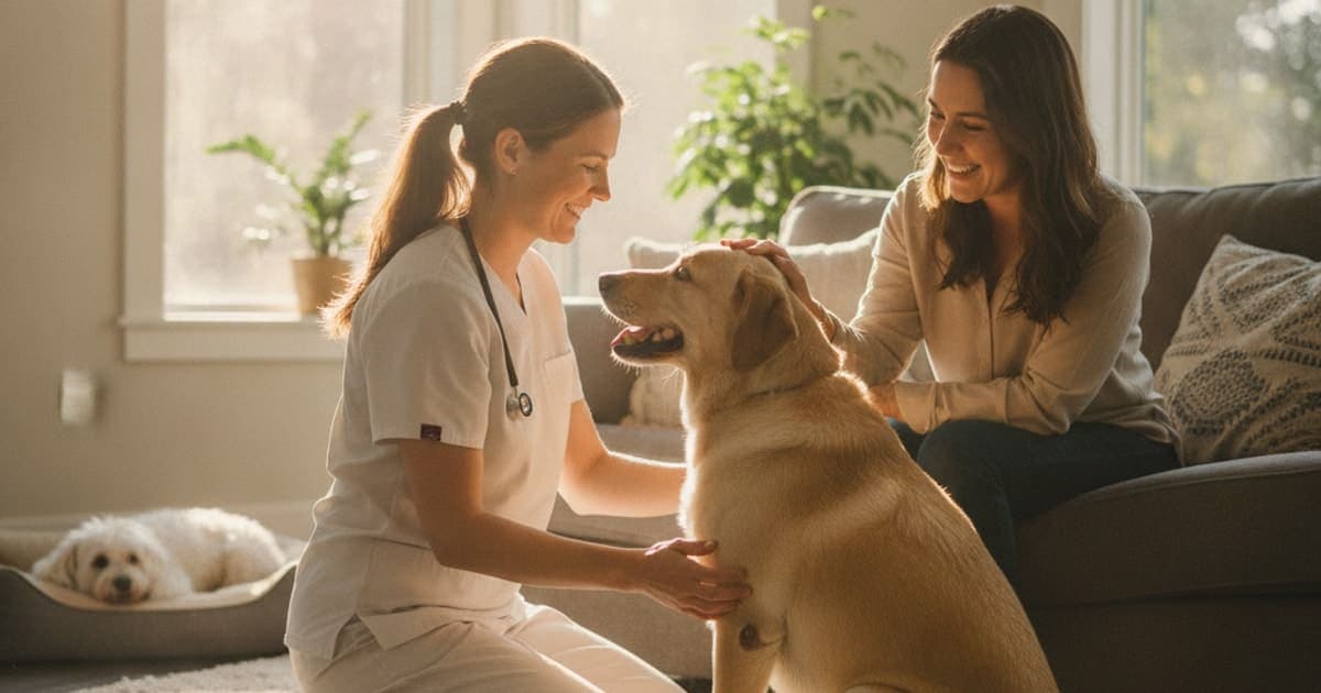
How Is Torn ACL Diagnosed in Dogs?
A torn ACL in dogs is diagnosed by a hands-on orthopedic exam of the knee — mainly the cranial drawer test and the tibial compression test — combined with watching the dog walk and X-rays of the joint. Sedation is often used so the leg muscles relax. The American College of Veterinary Surgeons describes this exam-plus-radiograph approach as the standard workup for cruciate disease.
Everything else — ultrasound, CT, MRI, arthroscopy — exists to answer questions the exam and X-rays leave open. Below is what each step actually looks like on the table, why sedation matters more than most owners expect, and what the diagnosis means for the weeks that follow.
The ACL in a dog is really the cranial cruciate ligament
Owners say ACL because that is the human term. In dogs the equivalent structure is the cranial cruciate ligament, or CCL. It runs diagonally through the stifle — the knee — and its job is to stop the tibia from sliding forward under the femur and to limit rotation of the joint.
The distinction matters for diagnosis, because canine cruciate injury usually does not happen the way a footballer's ACL tears. In dogs, the ligament more often degenerates over months or years, fraying under repeated load until a portion gives way. That is why so many owners describe a limp that came and went for weeks before the dramatic day their dog suddenly could not bear weight after a normal turn in the garden.
That degenerative pattern produces two diagnostic complications. First, partial tears are common, and a partially torn ligament can still resist the tests a veterinarian performs — so a subtle case can look normal to an inexperienced hand. Second, the meniscus, the cartilage cushion inside the joint, is frequently damaged alongside the ligament, and meniscal injury has its own signs to check for.
What your veterinarian is feeling for during the knee exam
The appointment starts before the dog is touched. Your vet will watch the dog stand, walk and trot, looking for which leg is unloaded, whether the toe barely touches down, and how the dog distributes weight when standing still. A dog with a cruciate problem often stands with the sore leg slightly forward and the toe tipped.
The sit test is another early clue. Dogs with an unstable or painful stifle often cannot fold the knee fully, so they swing the leg out to one side when they sit rather than tucking it neatly underneath.
Then the hands go on. Your vet will feel the thigh for muscle loss, which suggests the leg has been underused for weeks rather than days, and feel along the inside of the joint for medial buttress — a firm thickening that develops as the body tries to stabilise a loose knee. The joint capsule may feel swollen and puffy compared with the opposite leg.
Two specific tests carry most of the diagnostic weight:
- Cranial drawer test. The vet stabilises the femur with one hand and the tibia with the other, then attempts to slide the tibia forward. Any forward movement — the drawer sign — indicates the cranial cruciate ligament is not doing its job.
- Tibial compression test. The vet flexes the hock while holding the stifle steady, mimicking the load of weight-bearing. If the ligament is compromised, the tibia thrusts forward under the femur.
Both knees are always compared, because some dogs have naturally loose joints and because cruciate disease frequently affects the opposite leg too.
A confident cruciate diagnosis is made with the hands first — imaging confirms the picture and rules out other problems, but it is the drawer and tibial compression tests that make the call.
This is also where sedation earns its place. A tense, sore or simply nervous dog can brace the thigh muscles hard enough to mask several millimetres of instability, producing a false negative. Under light sedation the leg goes quiet, the tests become reliable, and clean X-rays can be taken in the same session. If your vet suggests sedating your dog for the exam, that recommendation is about accuracy, not convenience.
What X-rays show — and what they cannot
Radiographs do not show the cruciate ligament. Ligaments are soft tissue and simply do not appear on plain film. What X-rays show is the consequence of the injury and the state of the joint around it.
On a well-positioned lateral view of the stifle, a vet looks for joint effusion — fluid swelling that displaces the infrapatellar fat pad — as well as osteophytes, the small bony spurs that build up along the joint margins once the knee has been unstable for a while. Osteophytes are useful because they hint at how long the problem has been developing, which shapes the conversation about treatment.
X-rays also serve a second, non-negotiable purpose: ruling other things out. A hind-limb lameness in a large-breed dog can be caused by a bone tumour, and no responsible workup skips imaging that would reveal one. Radiographs also flag hip changes, fractures and other bone disease that could explain the limp.
If surgery is on the table, imaging becomes planning. For a tibial plateau levelling osteotomy, the surgeon measures the tibial plateau angle from a precisely positioned radiograph to calculate how the bone will be rotated.
Comparing the tools used to reach a diagnosis
| Method | What it shows | Main limitation |
|---|---|---|
| Orthopedic exam (drawer, tibial compression) | Direct evidence of joint instability | Partial tears and tense dogs can mask instability |
| Sedated exam | Reliable joint laxity assessment, better positioning | Requires anaesthetic assessment and monitoring |
| Radiographs (X-ray) | Effusion, arthritis, bone disease, surgical measurements | Cannot visualise the ligament itself |
| Ultrasound | Soft tissue detail in some joints; operator-dependent | Limited view inside the stifle |
| CT / MRI | Detailed bone and soft tissue imaging | Cost, availability, general anaesthesia |
| Arthroscopy | Direct visual assessment of ligament and meniscus | Invasive; usually done alongside surgery |
Most dogs never need anything past the first three rows. Advanced imaging and arthroscopy come in when the exam is equivocal, when a partial tear is suspected but instability cannot be demonstrated, or when a surgeon wants to inspect the meniscus directly during a procedure.
Problems that can look like a cruciate tear
A hind-leg limp is not automatically a cruciate injury, and part of the diagnostic process is systematically excluding the alternatives. Your veterinarian will be considering:
- Patellar luxation, where the kneecap slips out of its groove — common in small breeds and sometimes present alongside cruciate disease
- Hip dysplasia or hip arthritis, which can produce a similar hind-limb gait change
- Iliopsoas or other soft-tissue strains, which hurt on stretch but leave the stifle stable
- Lumbosacral and other spinal problems affecting the hind limbs
- Bone tumours, panosteitis in young dogs, and fractures
- Foot, nail and pad injuries, which are missed more often than anyone likes to admit
This is why a full orthopedic and neurological examination beats fixating on the knee. Two problems can coexist, and a dog with arthritic hips can still tear a cruciate ligament.
What happens once the diagnosis is confirmed
Once a cruciate tear is identified, the conversation moves to stabilising the joint. Surgical options include tibial plateau levelling osteotomy, tibial tuberosity advancement and extracapsular suture techniques, and the right choice depends on your dog's size, age, activity level, tibial plateau angle and the state of the meniscus. Conservative management — strict activity restriction, weight control, prescribed pain relief and structured rehabilitation — is a legitimate path for some dogs, more often smaller ones and those with partial tears.
Whichever route your veterinarian recommends, the medical plan comes first. Prescribed anti-inflammatories and pain relief exist for good reason, and they should never be stopped or substituted without the prescribing vet's input. Rehabilitation — controlled leash walking, range-of-motion work, hydrotherapy where available — is what converts a stabilised joint into a functioning leg.
Recovery is also a tissue-remodelling problem. Ligament, joint capsule, muscle and, after surgery, bone all rebuild over weeks and months, and that is the window in which many owners look for additional support. It is also the window in which the supplement aisle is at its worst: kitchen-sink chews with a dozen ingredients at trace levels, proprietary blends that hide how little is inside, and labels that lean on marketing rather than analysis.
pawgen takes the opposite approach with K9-REPAIR, a BPC-157 and TB-500 formulation made for dogs, dosed by body weight, third-party tested with certificates of analysis, and shipped direct to the door with a 60-day money-back guarantee. Research on BPC-157 suggests it interacts with pathways involved in tendon, ligament and gut tissue repair, and work on TB-500, a synthetic form of a thymosin beta-4 fragment, indicates a role in cell migration and new blood vessel formation — the processes that supply healing tissue. These peptides are not FDA-approved veterinary drugs, and nothing here should be read as a promise of any outcome for your dog. What can be said is that this is emerging and promising science, and that it is the stack a growing number of owners choose to run alongside veterinary care through a recovery. Talk to your veterinarian about how any supplement fits your dog's specific plan, particularly around a surgery date.
Key Takeaways
- The diagnosis is made by hand: the cranial drawer test and the tibial compression test demonstrate the instability that defines a cruciate tear.
- Sedation is frequently needed, because a tense dog can brace hard enough to hide joint laxity and create a false negative.
- X-rays cannot show the ligament. They show effusion, arthritis and bone disease, and they rule out serious alternatives such as tumours.
- Canine cruciate injury is usually degenerative, so partial tears and intermittent limping are common, and the second knee is often at risk.
- Advanced imaging and arthroscopy are reserved for unclear cases or for direct assessment of the meniscus.
- Once the diagnosis is confirmed, the surgical or conservative plan, pain control and structured rehab are the foundation of recovery.
A torn cruciate ligament is your veterinarian's call to make and their plan to direct — the exam, the imaging, the choice between surgery and conservative management, and the pain protocol all belong in their hands. Supplements and peptides operate in the space that proper veterinary care creates, supporting a dog through a recovery that a good diagnosis and a good plan have already set up.
For deeper reading, pawgen's guide to torn acl covers the condition end to end, with breed-specific recovery articles including the best supplement for collies with torn acl, the best supplement for briards with torn acl and the best supplement for kangals with torn acl. Owners dealing with skin issues during crate rest often read what are the first signs of hot spots in dogs and how much does it cost to treat hot spots in dogs, and full details on K9-REPAIR are on the main site.
Owners exploring peptide support for their dog can review K9-REPAIR — BPC-157 + TB-500 formulated for dogs — at https://pawgen.com/. BPC-157 and TB-500 are not FDA-approved veterinary drugs. Nothing in this article treats, cures or prevents any condition; every decision about your dog's care belongs with your veterinarian.
Frequently asked questions
- How much does it cost to treat torn ACL in dogs?
- Costs vary widely by region, clinic, dog size and the technique chosen. Surgical stabilisation is usually the largest expense, with osteotomy procedures generally costing more than extracapsular suture repair, plus diagnostics, anaesthesia, medication and rehabilitation. Ask your veterinary practice for a written estimate covering the whole recovery, not just the operation.
- Can a dog's torn ACL heal without surgery?
- The torn ligament itself does not reattach or regrow. Some dogs, particularly smaller ones and those with partial tears, stabilise acceptably with conservative management: strict activity restriction, weight control, prescribed pain relief and structured rehabilitation. Research suggests larger, more active dogs more often need surgical stabilisation to regain reliable limb function.
- What are the first signs of torn ACL in dogs?
- Sudden hind-limb lameness after a turn or jump, holding the leg up, toe-touching, stiffness after rest, sitting with the leg kicked out to one side, and reluctance to jump or climb stairs. Some dogs limp intermittently for weeks as a partial tear progresses. Any new hind-leg limp warrants a veterinary exam.
- What helps a dog with torn ACL?
- Veterinary care leads: an accurate diagnosis, a decision on surgery versus conservative management, prescribed pain control, weight management and structured rehabilitation such as controlled leash walking and hydrotherapy. Alongside that plan, many owners add recovery support like K9-REPAIR, a weight-dosed BPC-157 and TB-500 formulation for dogs, after discussing it with their vet.
- How long does torn ACL take to heal in dogs?
- After surgical stabilisation, dogs typically go through roughly eight to twelve weeks of strictly restricted activity, with a return to full function often taking several months as bone, ligament and muscle remodel. Conservative management is generally slower and less predictable. Follow your surgeon's specific rehab timeline rather than a general estimate.
- Is torn ACL in dogs painful?
- Yes. A cruciate tear causes joint instability, inflammation and often meniscal damage, all of which hurt. Dogs commonly mask pain, showing it as reduced activity, panting, restlessness, reluctance to jump or irritability rather than vocalising. Prescribed pain relief from your veterinarian is an essential part of the treatment plan.
- Does my dog need sedation for the knee exam?
- Often, yes. A tense or painful dog can brace the thigh muscles hard enough to hide several millimetres of instability, producing a false negative on the drawer test. Light sedation relaxes the limb, makes the cranial drawer and tibial compression tests reliable, and allows properly positioned X-rays in the same visit.
- Can a dog tear the cruciate ligament in both knees?
- Yes. Canine cruciate disease is frequently degenerative rather than a single traumatic event, so dogs that tear one cranial cruciate ligament often develop problems in the opposite knee later. Ask your veterinarian to assess both stifles at the time of diagnosis and to recheck the other knee at follow-up visits.
Educational content. BPC-157 and TB-500 are not FDA-approved veterinary drugs. Talk to your veterinarian before starting anything new, especially if your dog is on prescribed medication.