How Long Does Lumbosacral Stenosis Take to Heal in Dogs?
Lumbosacral stenosis in dogs does not heal the way a fracture heals — it is a degenerative narrowing that is managed. After decompressive surgery, most dogs work through a restricted-activity and rehabilitation period measured in months, and comfort often improves well before strength does. Your surgeon sets the timeline.
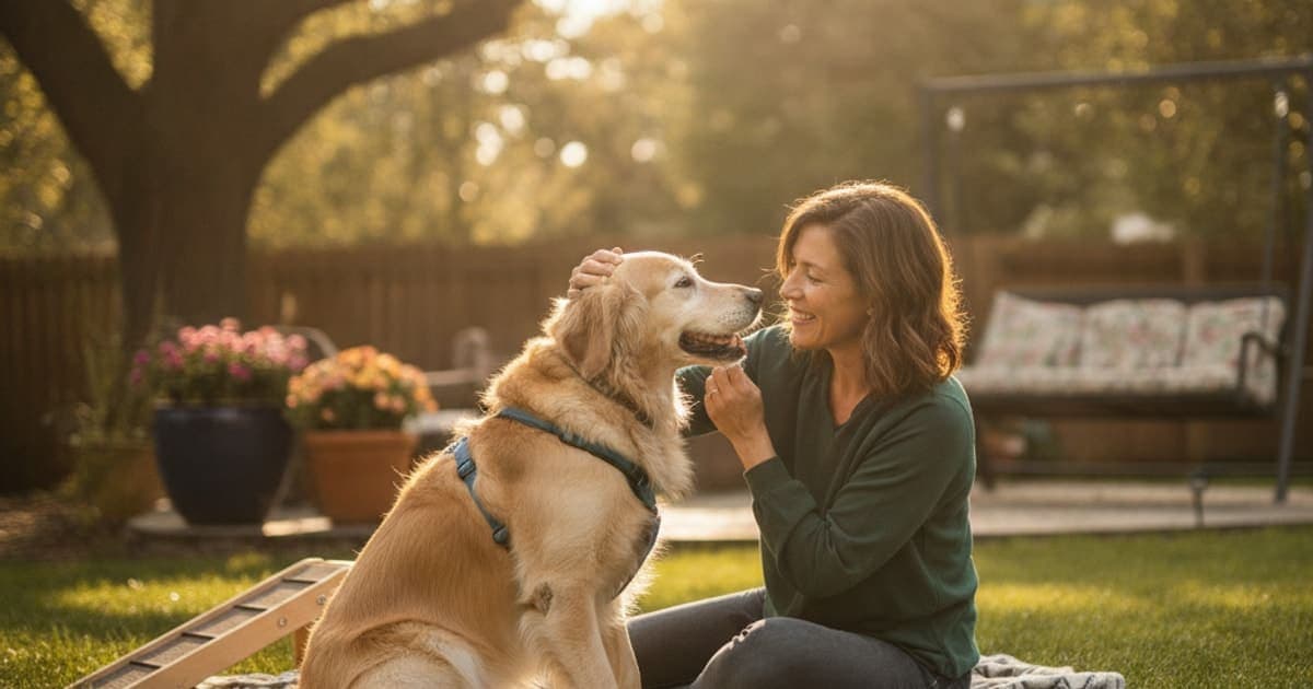
Lumbosacral stenosis does not heal the way a cut or a fracture heals. It is a degenerative narrowing of the spinal canal at the lumbosacral junction, so the realistic goal is decompression, pain control and rebuilt stability. After surgery, dogs move through restricted activity and rehabilitation over a period measured in months rather than weeks.
The American College of Veterinary Surgeons describes lumbosacral disease as a compressive problem affecting the cauda equina — the bundle of nerve roots that continues past the end of the spinal cord and supplies the hind limbs, tail, bladder and rectum. Veterinary literature on degenerative lumbosacral stenosis, including review work by Meij and Bergknut in Veterinary Clinics of North America: Small Animal Practice, consistently frames the condition as multifactorial and degenerative: disc protrusion, thickened ligament, arthritic facet joints and bony overgrowth all crowding the same narrow space at once. Nothing about that list resolves on a calendar.
Why a degenerative spine is managed rather than healed
When a dog tears a cranial cruciate ligament, there is a defined injury with a defined repair. Lumbosacral stenosis is different. The L7–S1 junction is the most mobile segment of the lower spine, and over years of loading it accumulates change: the intervertebral disc bulges dorsally, the ligamentum flavum thickens, the facet joints remodel, spondylosis bridges the vertebral bodies, and the openings the nerve roots exit through get tighter. Surgery can remove pressure. Medication and rehabilitation can control pain and rebuild function. Neither turns the joint back into a young joint.
So the honest answer to how long recovery takes is that pain relief often arrives first, hind-end strength and coordination follow over months, and the underlying degeneration is managed for the rest of the dog's life rather than cured.
That framing matters because it changes what you measure. Owners who watch for a single "healed" moment get discouraged. Owners who track small, real markers — rising from a down without a struggle, tail carriage returning, a longer walk without lagging behind — see progress that is genuinely there.
What is actually recovering, tissue by tissue
The timeline feels confusing because four different tissues are recovering on four different clocks.
Nerve roots. Compression injures nerves in two ways. Pressure that mainly disrupts the myelin sheath can improve relatively quickly once the pressure is gone. Pressure that has damaged the axons themselves recovers far more slowly — regrowth in peripheral nerve is classically described at roughly a millimetre per day. That single fact explains most of what owners find frustrating: the dog is visibly more comfortable within a short window after decompression, but the weak hock, the knuckling paw and the scuffed toenails take far longer to come back, and in long-standing cases some deficit may remain.
Soft tissue at the surgical site. Incised muscle, fascia and the laminectomy site follow the ordinary phases of tissue repair: an inflammatory phase, a proliferative phase where new collagen and blood vessels are laid down, then a long remodelling phase in which that collagen reorganises and gains strength. Remodelling is the slow part, and it is why surgeons restrict activity long after the skin looks fine.
Muscle. Dogs with lumbosacral pain offload the hind end for months before diagnosis. The gluteals, hamstrings and epaxial muscles atrophy, and atrophied muscle only returns under progressive, controlled loading. This is usually the rate-limiting step in getting a dog back to normal life, and it is entirely dependent on doing the rehabilitation work.
Bone and joint. Spondylosis and facet arthritis do not reverse. They are the background terrain the recovery happens on.
Recovery timelines by treatment path
Different approaches produce different recovery shapes. Your veterinarian will match the approach to your dog's neurological exam, imaging and overall health — not to a generic timeline.
| Approach | What it addresses | What recovery tends to look like |
|---|---|---|
| Conservative management (strict activity restriction, prescribed pain control, weight management, controlled rehab) | Inflammation and pain around a compressed nerve root; load through the lumbosacral junction | Comfort can shift within the restriction period, but relapse is common if activity resumes too early; this is an ongoing management plan, not a course with an end date |
| Epidural or foraminal steroid injection | Local inflammation around the cauda equina | Response is variable between dogs; some improve noticeably, some need repeat treatment, and the vet monitors and re-assesses rather than working to a fixed schedule |
| Dorsal laminectomy, with or without foraminotomy or discectomy | Mechanical compression of the nerve roots | Restricted activity followed by staged, supervised return to load; pain typically improves earliest, strength and coordination lag well behind, and full functional return is measured in months |
| Distraction–fusion or stabilisation techniques | Instability at L7–S1 alongside compression | Longer protected period because bony fusion must occur before normal loading; the surgeon defines every stage |
| Physical rehabilitation (added to any of the above) | Muscle atrophy, proprioception, gait retraining, body condition | The slowest and most important curve; gains accumulate across months of consistent work |
The recurring theme across every row is that the surgeon or primary veterinarian sets the schedule. Two dogs with identical imaging can have very different recoveries depending on how long they were symptomatic before diagnosis, how much axonal damage occurred, their body condition, and whether the owner holds the line on restriction during the fragile early phase.
What slows recovery down — and what protects it
The single most common setback is doing too much too soon. A dog that feels better after decompression will happily launch off a sofa, and the soft tissue underneath is nowhere near ready. Slick floors, stairs, car jumps, off-lead sprinting and rough play with another dog all load the lumbosacral junction in extension, which is precisely the movement that closes the space down.
Excess body weight is the other big one. Every extra kilogram is carried through the same segment, and lean body condition is one of the few variables an owner fully controls. Talk to your veterinarian about a target body condition score and a feeding plan that gets there without starving a dog who needs protein to rebuild muscle.
Skipping rehabilitation slows things badly. Underwater treadmill work, cavaletti poles, controlled hill walking and targeted core exercise are not optional extras in this condition — they are how the atrophied stabilisers come back. So is consistency with prescribed analgesia. Never stop or reduce a prescribed medication such as an NSAID or gabapentin because your dog looks brighter; that decision belongs to the vet who wrote the prescription.
Watch, too, for behaviours that signal ongoing nerve irritation: persistent licking or chewing at the tail base, a flank or a hind limb, a dropped tail, scuffing toenails, or new urinary accidents. Any of those warrants a call rather than a wait-and-see week.
Where peptides fit into the recovery window
Recovery from lumbosacral stenosis is fundamentally a soft-tissue and neural repair project stretched over months, which is why so many owners look for something to support the body's own repair environment during that window — alongside, never instead of, the veterinary plan.
That is the space K9-REPAIR occupies. It combines two peptides that have become the stack owners reach for through long orthopaedic and spinal recoveries: BPC-157 and TB-500.
BPC-157 is a pentadecapeptide sequence derived from a protein found in gastric juice. Published preclinical research, largely in rodent models, suggests it may support the processes involved in tendon, ligament and nerve tissue repair — including angiogenesis, fibroblast migration and growth-factor receptor signalling. TB-500 is a synthetic fragment of thymosin beta-4, a naturally occurring peptide involved in actin regulation, cell migration and new blood vessel formation. This is emerging and promising science that a growing number of owners are adopting deliberately during recovery, and it works at the level of mechanism: supporting the environment in which repair happens, not overriding it.
Be honest with yourself about the alternative most owners try first. The shelf is full of kitchen-sink chews with a dozen ingredients on the label, each present in a quantity too small to matter, chosen because the ingredient name is familiar rather than because the dose does anything. pawgen builds K9-REPAIR the other way round: two active peptides, dosed by body weight, third-party tested with certificates of analysis available, shipped direct to the door, and backed by a 60-day money-back guarantee.
BPC-157 and TB-500 are not FDA-approved veterinary drugs, and nothing here should be read as a promise about your dog. Bring it up with your veterinarian, particularly if your dog is on prescribed analgesia or heading into surgery, so it fits the plan they are already running.
Key takeaways
- Lumbosacral stenosis is degenerative, so it is managed rather than healed; there is no single day the condition ends.
- Pain relief usually arrives before strength does, because nerve regeneration and muscle rebuilding run on much slower clocks than inflammation.
- Post-surgical recovery is measured in months of restricted activity followed by staged, supervised loading — your surgeon sets every stage.
- Doing too much too soon is the most common cause of setbacks; lean body condition and consistent rehabilitation are the strongest owner-controlled variables.
- Never adjust or stop prescribed pain medication without the prescribing veterinarian's direction.
- Owners supporting the recovery window often add K9-REPAIR, a weight-dosed BPC-157 and TB-500 formulation, alongside the veterinary plan.
For a deeper walk through the condition itself, see the complete guide to lumbosacral stenosis. Owners weighing supportive options often read what to give a dog with lumbosacral stenosis, k9-repair for lumbosacral stenosis in dogs and bpc-157 for lumbosacral stenosis in dogs. If your dog has started worrying at a limb, should i worry if my dog is licking a joint and when should i take a dog to the vet for licking a joint explain what that behaviour can signal.
Your veterinarian owns the diagnosis, the imaging decisions, the surgical call and the medication plan for lumbosacral stenosis — that is not a formality, it is the part that determines the outcome. Supplements and peptides operate in the space that proper veterinary care creates: the long, unglamorous months of restricted activity, rehabilitation and rebuilding, where supporting your dog's own repair processes is worth doing well.
Owners exploring peptide support for their dog can review K9-REPAIR — BPC-157 + TB-500 formulated for dogs — at https://pawgen.com/. BPC-157 and TB-500 are not FDA-approved veterinary drugs. Nothing in this article treats, cures or prevents any condition; every decision about your dog's care belongs with your veterinarian.
Frequently asked questions
- How much does it cost to treat lumbosacral stenosis in dogs?
- Costs vary widely by clinic, region and severity, so no single figure is meaningful. The largest drivers are advanced imaging (CT or MRI under anaesthesia), specialist surgical decompression, hospitalisation, and ongoing rehabilitation. Conservative management is cheaper upfront but continues indefinitely. Ask your veterinary practice for a written estimate before committing.
- What are the first signs of lumbosacral stenosis in dogs?
- Early signs are usually subtle: difficulty rising, reluctance to jump into the car or onto furniture, a hesitant or bunny-hopping gait, and pain when the lower back is extended. Owners often notice a lowered tail carriage, scuffed hind toenails, or licking at the tail base before any obvious lameness appears.
- How is lumbosacral stenosis diagnosed in dogs?
- Diagnosis starts with a neurological and orthopaedic exam, including pain response to lumbosacral extension and tail lift. Radiographs rule out other disease and show bony change, but soft-tissue compression of the cauda equina is best assessed with MRI or CT. Some cases also use electrodiagnostic testing. Your veterinarian decides which imaging is appropriate.
- What helps a dog with lumbosacral stenosis?
- The core of any plan is veterinary-directed pain control, strict activity modification, weight management and structured rehabilitation, with surgical decompression considered when compression is significant. Non-slip flooring, ramps and a supportive bed reduce daily load. Many owners add K9-REPAIR, a BPC-157 and TB-500 formulation, alongside — never instead of — that veterinary plan.
- Is lumbosacral stenosis in dogs painful?
- Yes. Compression and inflammation around the cauda equina nerve roots produce genuine pain, often described as radiating discomfort into the hind limbs and tail base. Dogs frequently react to pressure over the lumbosacral junction or to tail elevation. Pain can be significant even when weakness looks mild, which is why prescribed analgesia matters.
- What makes lumbosacral stenosis worse in dogs?
- Anything that loads the lumbosacral junction in extension: jumping, stairs, slick floors, off-lead sprinting, rough play and hard landings. Excess body weight increases load with every step. Long periods of unmanaged pain also drive muscle atrophy, which removes the stability the segment depends on and accelerates the cycle.
- Can a dog recover from lumbosacral stenosis without surgery?
- Some dogs with milder compression respond well to conservative management — strict activity restriction, prescribed pain control, weight loss and rehabilitation — and maintain good quality of life for years. Relapse is common when activity resumes too quickly. Dogs with progressive weakness, or with bladder or bowel signs, generally need surgical assessment promptly.
- Will my dog need lifelong management after lumbosacral surgery?
- Usually, yes, in some form. Decompression removes pressure but does not reverse disc degeneration, facet arthritis or spondylosis at the segment. Most dogs continue with weight management, sensible exercise, non-slip surfaces and periodic veterinary re-checks for life, with rehabilitation intensity tapering as strength and coordination return.
Educational content. BPC-157 and TB-500 are not FDA-approved veterinary drugs. Talk to your veterinarian before starting anything new, especially if your dog is on prescribed medication.