Megaesophagus in Dogs: X-Ray vs Normal Findings
Megaesophagus in dogs is diagnosed with chest radiographs, where a dilated esophagus shows up as a soft tissue or gas filled column that a normal film does not show. If plain x-rays are unclear, a vet may add a barium swallow study, endoscopy and bloodwork to look for an underlying cause.
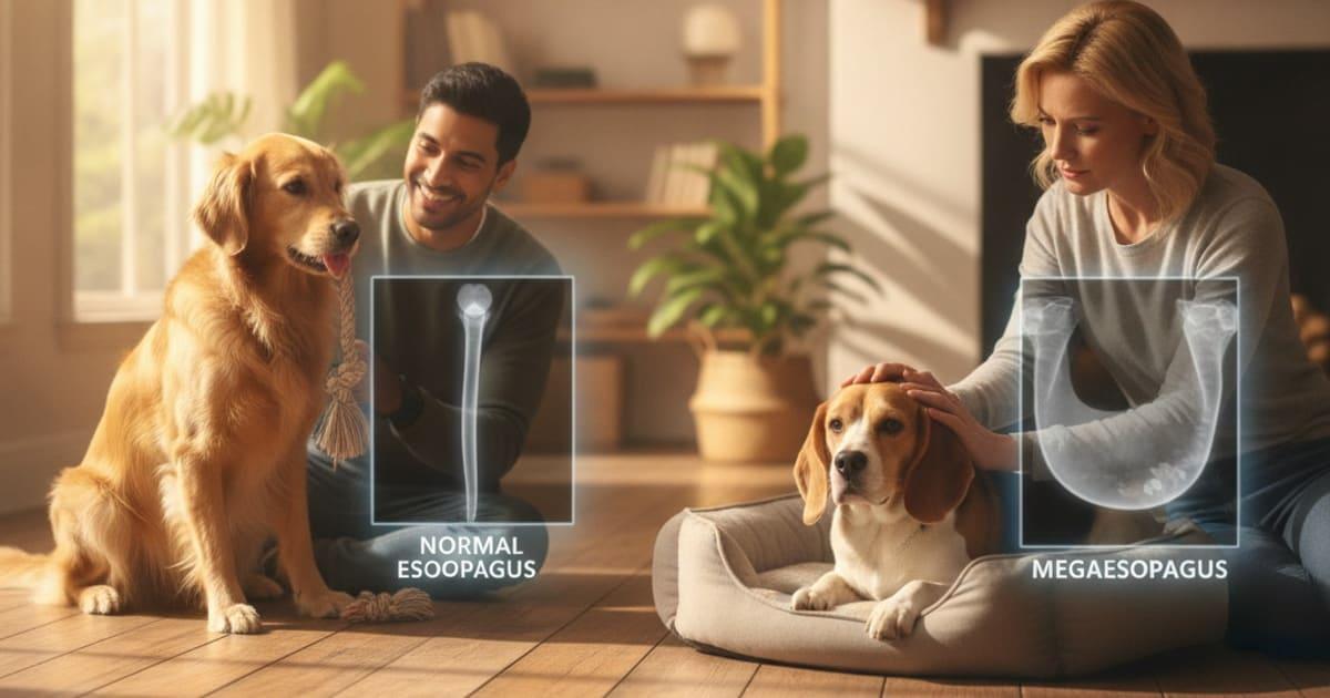
Megaesophagus in dogs is diagnosed with chest x-rays, and the call is made by comparing the film against what a normal chest should look like: a normal esophagus is essentially invisible on radiographs, while a megaesophagus shows up as a widened, air or food filled tube running down the chest. If your dog is bringing food back up and is also coughing, breathing fast or noisily, running a fever or going flat and quiet, call your veterinarian now or go to an emergency clinic, because those are the signs of aspiration pneumonia.
Aspiration pneumonia is the complication that turns megaesophagus from a feeding problem into an emergency, so breathing changes in a dog that regurgitates always outrank waiting for an appointment.
The rest of the workup builds outward from that first set of radiographs. If plain films are not conclusive, a veterinarian may add a barium swallow study, and separately may recommend endoscopy and blood testing, not to confirm the dilation but to hunt for what is causing it.
Regurgitation and vomiting are not the same thing, and the difference matters
The single most useful thing an owner brings to the appointment is an accurate description of what came up and how.
Vomiting is active. There is nausea first, drooling, lip licking, restlessness, then heaving abdominal contractions. What comes up is usually partly digested and may contain bile.
Regurgitation is passive. The dog lowers its head and material falls out, sometimes with no warning at all, sometimes minutes after eating and sometimes much later. The food often looks undigested, may be tube shaped, and is frequently coated in foamy saliva.
That distinction steers the whole workup. Vomiting sends a veterinarian looking at the stomach, intestines, pancreas, kidneys and liver. Regurgitation sends them looking at the esophagus, and the esophagus is examined with imaging. A phone video of an episode is genuinely useful evidence, as is a note of whether the dog eats standing, how fast it eats, and whether episodes cluster around certain foods or textures.
Other details worth writing down before the visit: weight loss despite a good appetite, ravenous hunger, bad breath, a wet or productive sounding cough, nasal discharge, voice change, muscle weakness that gets worse with exercise, or a puppy that stopped thriving right when it moved onto solid food.
What a dilated esophagus looks like on radiographs compared with a normal chest
Radiographs, often shortened to rads, are the starting point. Most of the time plain films are enough to identify a markedly dilated esophagus.
On a normal chest, the esophagus is a collapsed, empty tube sitting between the trachea and the spine. It contains no air column and casts no distinct shadow, so you simply do not see it. The spine, heart, trachea and lung fields are what stand out.
On a chest with megaesophagus, the esophagus becomes visible because it is holding something: swallowed air, fluid, saliva or food. That gives the radiologist a structure to see where there should be nothing.
| Feature | Normal chest radiograph | Pattern seen with megaesophagus |
|---|---|---|
| Esophagus itself | Not visible as a distinct structure | Visible as a widened tube running from the thoracic inlet toward the diaphragm |
| Contents | None | Gas, fluid, saliva or food material filling the tube |
| Trachea | Clearly outlined, sitting in its usual position | May be pushed downward, and the wall between trachea and esophagus can appear as a single thickened stripe |
| Border near the diaphragm | Clean, uninterrupted outline | Converging soft tissue lines can be seen where the dilated esophagus narrows at the diaphragm |
| VD or DV view | Esophagus not apparent | The dilated structure can be seen widening to either side of the midline |
| Lung fields | Evenly aerated | May show patchy changes in the dependent lobes if aspiration has occurred |
There is no owner-usable number here. Radiologists interpret esophageal width in context, comparing it against neighbouring landmarks such as the vertebral bodies, the trachea and the heart, and weighing that against the dog's size, positioning and breathing phase. That judgement is what you are paying for, and it is not something to attempt from a phone photo of a film.
Why one view is never enough
A single lateral x-ray can miss or exaggerate a dilated esophagus, so a veterinarian will usually take a set.
A standard study includes both a left and right lateral view plus a ventrodorsal or dorsoventral view. The VD view, taken with the dog on its back, lets the reader see the esophagus spreading on both sides of the midline and separates esophageal gas from gas sitting inside the trachea or stomach. Comparing the two laterals also helps, because gravity shifts fluid and gas between lung lobes and between segments of the esophagus depending on which side the dog is lying on.
There is a practical catch worth knowing about. A dog that is panting, frightened, gulping air or lightly sedated can swallow enough air to produce a transient gas pocket in the esophagus that is not true megaesophagus. Equally, a dog with real megaesophagus can produce a near normal looking film if the esophagus happened to be empty when the picture was taken. That is why a veterinarian may repeat imaging, change positioning, or move on to a contrast study rather than closing the case on one picture.
If aspiration is suspected, the same films are also read for pneumonia, since that finding changes what needs doing first.
When a barium swallow study is added
A barium study is the next step when plain films are equivocal, when the clinical picture is strongly suggestive but the radiographs are not, or when the question is how the esophagus moves rather than how wide it is.
Barium is a contrast liquid that shows up brightly on x-ray. The dog swallows it, sometimes mixed with food of different textures, and further images are taken. The contrast outlines the inside of the esophagus, so a dilated segment, a narrowed segment or a pocket where material pools becomes obvious.
A fluoroscopic swallow study goes further. Fluoroscopy is live, moving x-ray, so instead of a frozen moment the clinician watches the swallow happen: whether the wave of muscle contraction travels normally, whether material stalls, whether it moves backward, and whether anything spills toward the airway. That motion information is something still radiographs cannot provide, and it is why referral for fluoroscopy is sometimes recommended.
Barium is not risk free in a dog that is already regurgitating, because contrast can be aspirated. Your veterinarian weighs that against the value of the information, and may hold off entirely in a dog with active pneumonia. This is a decision to make with them, not a test to request as a default.
Endoscopy and the search for an underlying cause
Confirming a dilated esophagus is only half the job. The more important question is usually why, because the cause is what the treatment plan is built around.
Endoscopy puts a camera into the esophagus under general anaesthesia. It is less about measuring dilation and more about looking at the lining and at anything blocking the path: inflammation from reflux, a stricture, a retained foreign object, a mass, or a hiatal hernia sliding up through the diaphragm. Biopsies can be taken during the same procedure.
Alongside imaging, a veterinarian may run blood and urine testing to screen for systemic illness, hormonal disease, and inflammation, and may recommend specific tests for neuromuscular disease, including antibody testing for myasthenia gravis, which is one of the recognised causes of acquired esophageal dilation in adult dogs. Depending on findings, chest CT or further neurological assessment may follow.
Some cases never yield an identifiable cause and are classed as idiopathic. That is a legitimate diagnosis reached after a proper search, not a shrug.
What else can look like this on a first-pass film
Part of diagnosis is ruling things out. A veterinarian reading a chest film in a regurgitating dog is also weighing:
- Esophagitis and reflux, which can cause regurgitation with little or no visible dilation
- Esophageal stricture, a narrowed scarred segment that backs material up behind it
- A swallowed foreign object lodged in the esophagus
- Vascular ring anomaly, where a band of vessel tissue constricts the esophagus near the heart base, classically producing dilation in front of the heart in a young dog
- Hiatal hernia, which may come and go depending on when the image is taken
- Pharyngeal or laryngeal swallowing disorders, where the problem is above the esophagus
- Transient air swallowing from stress, sedation or respiratory distress
- Aspiration pneumonia, which can be both a consequence and the reason the dog was brought in
Each of these is distinguished by a different combination of views, contrast, endoscopy and bloodwork, which is exactly why the workup is staged rather than done all at once.
Key takeaways
- A normal esophagus is invisible on chest x-rays. Megaesophagus is diagnosed when a widened, gas, fluid or food filled esophagus becomes visible where nothing should be seen.
- Multiple views, including both laterals and a VD or DV, reduce the chance of a false impression in either direction.
- Esophageal width is interpreted against anatomical landmarks by a trained reader. There is no number for owners to apply at home.
- A barium swallow or fluoroscopic study is added when plain films are unclear or when swallowing motion needs to be assessed, and carries its own aspiration risk to weigh with your vet.
- Endoscopy and blood testing are aimed at finding a cause rather than confirming the dilation.
- Coughing, fast or noisy breathing, fever or lethargy in a regurgitating dog points toward aspiration pneumonia and needs same-day veterinary attention.
Working through this with your veterinarian
Bring the history, bring a video if you have one, and describe episodes precisely. Ask which views were taken, whether contrast is being considered, and what the plan is for identifying an underlying cause. Results vary from dog to dog, so follow-up imaging and testing are planned around your individual dog rather than a fixed template. If anything about breathing changes between appointments, talk to your vet straight away rather than waiting for the next scheduled visit.
If you are unsure whether what you are seeing tonight counts as an emergency, Dog Emergency Signs sets out the red flags worth acting on immediately. Your veterinarian owns the diagnosis and the treatment plan here: the imaging, the contrast study, the interpretation and everything that follows belong with them, and the most useful thing you can do is get your dog in front of them early with a clear account of what you have seen.
Frequently asked questions
- How is megaesophagus in dogs diagnosed?
- It is diagnosed with chest radiographs. A veterinarian looks for a widened esophagus filled with gas, fluid or food, where a normal esophagus would not be visible at all. If plain films are unclear, a barium swallow study may be added, along with bloodwork and endoscopy to identify an underlying cause.
- How to diagnose megaesophagus in dogs?
- Diagnosis belongs with a veterinarian and begins with a set of chest x-rays taken in several positions, including a VD or DV view. Contrast imaging with barium, or live fluoroscopic swallow imaging, may follow. Further testing looks for causes such as neuromuscular, hormonal or structural disease.
- How to test for megaesophagus in dogs?
- The core test is plain chest radiography, usually both lateral views plus a ventrodorsal view. A barium swallow study can outline the inside of the esophagus when plain films are equivocal. Endoscopy and blood testing are added to search for a cause rather than to confirm the dilation itself.
- How to check for megaesophagus in dogs?
- You cannot check for it at home, but you can gather what the vet needs: note whether material comes up passively without heaving, how long after eating, and whether food looks undigested. Film an episode if you can. Imaging at the clinic is what confirms or rules it out.
- What does megaesophagus look like on a dog x-ray?
- It appears as a widened tube running from the thoracic inlet toward the diaphragm, filled with gas, fluid or food. The trachea may be displaced downward, the wall between trachea and esophagus can look like a single thickened stripe, and converging soft tissue lines may appear near the diaphragm.
- Can megaesophagus be missed on an x-ray?
- Yes. If the esophagus happens to be empty when the image is taken, the film can look close to normal. The reverse also happens, since a panting, stressed or sedated dog may swallow air and create a temporary gas pocket. Repeat or contrast imaging resolves the uncertainty.
- What is a barium study for megaesophagus in dogs?
- The dog swallows a contrast liquid that shows brightly on x-ray, sometimes mixed with foods of different textures. Follow-up images show the inside outline of the esophagus, revealing dilated segments, narrowings or pooling. Barium carries aspiration risk in a regurgitating dog, so the decision is made with your veterinarian.
- Is endoscopy needed to diagnose megaesophagus in dogs?
- Not usually for the dilation itself, which radiographs generally show. Endoscopy is recommended when the lining needs direct inspection or an obstruction is suspected, looking for inflammation, a stricture, a foreign object, a mass or a hiatal hernia. It is performed under general anaesthesia and allows biopsies.
- Can megaesophagus be diagnosed in a puppy on x-ray?
- Yes, and it is often picked up around weaning when solid food is introduced and a puppy starts regurgitating or fails to thrive. The same radiographic approach applies. In young dogs, a veterinarian also considers a vascular ring anomaly constricting the esophagus near the heart base.
- When does regurgitation become an emergency?
- When breathing is involved. Coughing, fast or laboured breathing, noisy or wet sounding respiration, blue or pale gums, fever, collapse or sudden lethargy in a dog that regurgitates can indicate aspiration pneumonia. That warrants immediate veterinary attention rather than waiting for a routine appointment.
Educational content, not veterinary advice. Ask your vet about your dog.