How Is Hind End Weakness Diagnosed in Dogs? (Vet Workup)
Hind end weakness in dogs is diagnosed by combining a neurologic exam, an orthopedic exam and gait observation to localize the problem, then confirming it with imaging such as radiographs, CT or MRI plus bloodwork. The hands-on exam decides which tests are worth running, not the other way around.
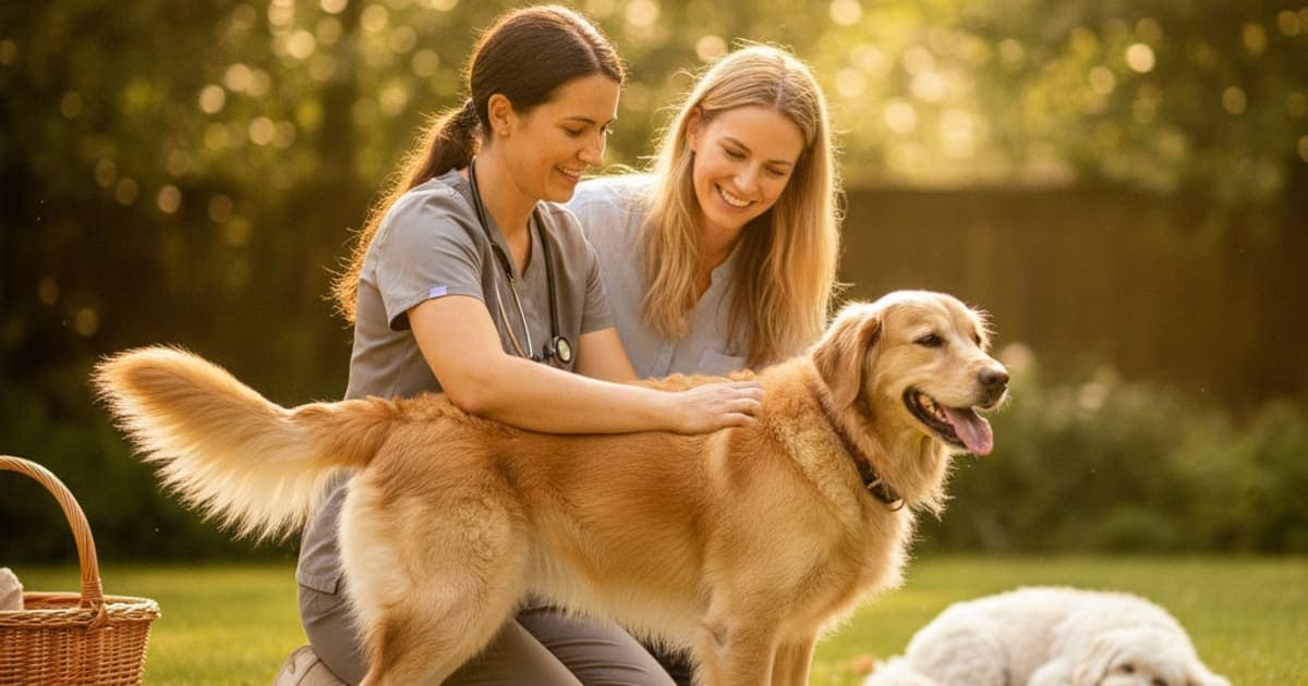
How Is Hind End Weakness Diagnosed in Dogs?
Hind end weakness in dogs is diagnosed by combining a full neurologic exam, an orthopedic exam and careful gait observation to localize the problem, then confirming it with imaging — radiographs, CT or MRI — plus bloodwork. The exam determines whether the cause sits in the spine, the joints, the muscles or the body as a whole, and imaging follows from there.
Weak back legs is a symptom, not a diagnosis. The same picture — a dog who struggles on stairs, sways at the hips, or sits down halfway through a walk — can come from a knee, a hip, a spinal disc, a nerve root, or a metabolic problem that has nothing to do with the legs at all. The workup exists to tell those apart, in a specific order, and understanding that order makes you a far more useful partner in the exam room.
Step one: nerves, joints, or the whole body?
Before any machine is switched on, your veterinarian is answering one question: which system is failing?
Neurologic weakness means the signal from the brain to the legs is being interrupted somewhere — a disc pressing on the spinal cord, a fibrocartilaginous embolism, lumbosacral nerve root compression, degenerative myelopathy. These dogs often scuff their nails, knuckle a paw over, cross their back legs, or have a wobbling, uncoordinated gait. Pain may or may not be present.
Orthopedic weakness means the hardware hurts. Cranial cruciate ligament disease is one of the most commonly diagnosed causes of hind limb lameness in dogs, and hip dysplasia and osteoarthritis are close behind. These dogs usually shift weight off one leg, are stiff after rest, sit with a leg kicked out to the side, and resent specific joint movements.
Systemic weakness means the legs are fine and the body is not — anemia, endocrine disease, tick-borne infection, cardiac disease, myasthenia gravis, or generalized muscle loss in an older dog. These dogs are typically weak everywhere, tire quickly, and get worse with exertion rather than with a particular movement.
Most of the diagnostic value comes from this first sort. A dog who knuckles gets a different test list than a dog who yelps when a knee is flexed.
What the neurologic exam is actually testing
The neurologic exam looks simple and is not. Each piece narrows the location of the problem along the spinal cord.
Proprioceptive placing. Your vet flips a back paw so the dog stands on its knuckles. A neurologically normal dog corrects it almost instantly. Delayed correction suggests the nerve pathways carrying position sense are compromised.
Spinal reflexes. Patellar and withdrawal reflexes help separate an upper motor neuron problem (lesion in the mid-back, reflexes normal to exaggerated) from a lower motor neuron problem (lesion in the lower lumbar spine or nerve roots, reflexes reduced). This is how a vet distinguishes a T3–L3 lesion from an L4–S3 lesion without an image.
Muscle mass and tone. Symmetry matters. Muscle loss on one side points toward chronic offloading of a painful limb; loss on both sides suggests nerve supply or systemic disease.
Spinal palpation. Running along the vertebral column looking for a focal pain response, and extending the hips and lumbosacral junction to check for the discomfort typical of cauda equina syndrome.
Tail, anal tone and bladder function. Involvement here signals a lesion at the very end of the spinal cord and raises urgency considerably.
The exam localizes the problem and the imaging only confirms it — which is why a veterinarian who watches your dog walk before laying a hand on them is doing the single most valuable part of the entire workup.
The orthopedic exam, including the tests for a torn cruciate
If the neurologic exam comes back clean, attention turns to joints. Your vet will palpate each hind limb joint through its range of motion, feeling for effusion, thickening, crepitus, reduced range and a pain response.
For the stifle (knee), the specific manoeuvres are the cranial drawer test and the tibial compression test, which check whether the tibia slides forward abnormally against the femur — the hallmark of cruciate instability. A medial buttress, or firm thickening on the inside of the knee, is a common finding in longer-standing cases. The sit test is telling too: dogs with knee pain often sit with the affected leg swung out rather than tucked underneath.
For the hips, the Ortolani test assesses laxity in younger dogs, and hip extension is checked for pain in older ones. Tense or painful dogs are frequently sedated for this part, because muscle guarding can mask instability entirely. Sedation also allows properly positioned radiographs in the same visit. If a cruciate problem is suspected, the detail in how is cruciate ligament rupture diagnosed in dogs covers that pathway in depth.
Imaging, bloodwork and DNA testing: what each one can and cannot show
No single test answers everything. Each rules a category in or out.
| Test | What it examines | What it helps rule in or out |
|---|---|---|
| Neurologic and orthopedic exam | Reflexes, proprioception, joint stability, pain response | Localizes the problem to a spinal segment, a joint or a systemic cause |
| Radiographs (X-rays) | Bone, joint spaces, joint effusion, spinal alignment | Hip dysplasia, osteoarthritis, fractures, bone tumours, narrowed disc spaces |
| CT | Cross-sectional bone and mineralized tissue detail | Lumbosacral disease, mineralized disc material, complex bony change |
| MRI | Spinal cord, discs, nerve roots, soft tissue | Intervertebral disc disease, FCE, spinal cord tumours, nerve root compression |
| Bloodwork and urinalysis | Organ function, inflammation, endocrine markers | Systemic and metabolic contributors to generalized weakness |
| Tick-borne disease panel | Infectious causes of joint and neurologic signs | Infection-driven polyarthritis or neurologic disease |
| Joint fluid analysis | Cells and fluid within the joint | Immune-mediated polyarthritis, septic arthritis |
| SOD1 cheek-swab DNA test | Genetic risk for degenerative myelopathy | Whether a dog carries the at-risk genotype |
On that last line: research led at the University of Missouri and published in the Proceedings of the National Academy of Sciences in 2009 identified a mutation in the SOD1 gene associated with canine degenerative myelopathy, which is why a simple cheek swab is now part of the workup for at-risk breeds such as German Shepherds, Boxers and Corgis. Importantly, the DNA result describes risk, not a confirmed diagnosis — degenerative myelopathy is a diagnosis of exclusion in the living dog, reached by ruling out compressive spinal disease on MRI.
Radiographs are usually the first imaging step because they are widely available and answer the orthopedic questions well. They do not show the spinal cord. If the exam points to a nerve problem, advanced imaging under general anaesthesia becomes the meaningful next step, and that conversation belongs with your veterinarian, who can weigh your dog's age, anaesthetic risk and the likely treatment paths.
How to help your vet reach the answer faster
Much of the diagnostic picture comes from the history you provide, and most dogs walk suspiciously well in the exam room.
Film your dog before the appointment. Take video from the side and from directly behind, on a non-slip surface, at a walk and at a slow trot. Include the moments you actually worry about: getting up from lying down, taking stairs, sitting, and jumping into the car. Note whether the difficulty appeared over hours, weeks or months; whether it is worse cold or after exercise; whether it affects one leg or both; and whether the nails on one paw are wearing down unevenly. Bring a list of every medication and supplement, including doses. If your dog is clearly painful, note where. The broader background on hind end weakness is worth reading before you go so you know what to describe.
Where recovery support fits once you have a diagnosis
A diagnosis sets the plan: surgery for an unstable stifle, prescribed pain control and rehabilitation for arthritis, decompression or strict rest for disc disease, physical therapy for weakness of any origin. That plan comes from your veterinarian, and nothing should replace it.
Within that plan, soft tissue still has to remodel. Ligament, tendon and joint capsule repair on their own timeline, driven by blood supply, collagen turnover and controlled loading — and that is the window many owners look to support. K9-REPAIR from pawgen combines BPC-157 and TB-500, two peptides owners have adopted through recovery periods. BPC-157 is a synthetic peptide based on a sequence identified in gastric juice, and research in animal models suggests it may support new blood vessel formation and tendon and ligament cell activity. TB-500 is related to thymosin beta-4, a naturally occurring protein involved in actin regulation and cell migration — the machinery cells use to move into an injured area. The science here is emerging and promising, and the products are not FDA-approved veterinary drugs; pawgen publishes third-party certificates of analysis, gives weight-based guidance, ships direct to your door and backs orders with a 60-day money-back guarantee. What it is not is a substitute for surgery, for a prescribed medication, or for the diagnosis itself.
Be skeptical of the other end of the shelf: kitchen-sink mobility chews with a dozen ingredients at token amounts, proprietary blends that hide quantities, and brands that spend more on packaging than on testing.
Key Takeaways
- Hind end weakness is a symptom with many causes; the diagnosis comes from localizing it to nerves, joints or a systemic problem.
- The neurologic exam — proprioception, reflexes, muscle symmetry, spinal palpation — determines which imaging is worth doing.
- Radiographs answer orthopedic questions; MRI is what shows the spinal cord, discs and nerve roots.
- Cruciate disease is checked with cranial drawer and tibial compression tests, often under sedation.
- A SOD1 cheek swab identifies genetic risk for degenerative myelopathy but does not confirm it in a living dog.
- Video of your dog walking, plus a precise timeline, meaningfully speeds up the workup.
For more on the questions owners ask next, see what helps a dog with hind end weakness, how long does hind end weakness take to heal in dogs, is hind end weakness in dogs painful, and what helps a dog with partial ccl tear.
Your veterinarian owns the diagnosis and the treatment plan — the exam, the imaging, the surgical decision and the prescriptions. Talk to your veterinarian before adding anything to a recovering dog's routine, particularly if that dog is on medication, is a puppy, or is pregnant or nursing. Supplements and peptides work only in the space that proper veterinary care creates.
Owners exploring peptide support for their dog can review K9-REPAIR — BPC-157 + TB-500 formulated for dogs — at https://pawgen.com/. BPC-157 and TB-500 are not FDA-approved veterinary drugs. Nothing in this article treats, cures or prevents any condition; every decision about your dog's care belongs with your veterinarian.
Frequently asked questions
- What helps a dog with hind end weakness?
- What helps depends entirely on the cause, so the diagnosis comes first. Common elements include veterinary-directed pain management, controlled exercise and physical rehabilitation, weight management, non-slip flooring, ramps, and surgery where a joint is unstable. Many owners also add recovery support such as K9-REPAIR alongside — never instead of — the plan their veterinarian set.
- How long does hind end weakness take to heal in dogs?
- Recovery time depends on the underlying cause. Soft tissue and post-surgical healing typically progresses over weeks to months as collagen remodels and muscle rebuilds, while degenerative conditions are managed rather than resolved. Your veterinarian can give a realistic timeline once imaging and exam findings identify what is actually driving the weakness.
- Is hind end weakness in dogs painful?
- Sometimes, and sometimes not. Orthopedic causes such as cruciate disease, hip dysplasia and arthritis are usually painful. Certain neurologic causes, including degenerative myelopathy and fibrocartilaginous embolism, often are not. Dogs mask pain well, so panting, reluctance to be touched, restlessness, licking a joint or changed sleeping posture matter more than vocalizing.
- What makes hind end weakness worse in dogs?
- Slippery floors, stairs, jumping on and off furniture, excess body weight, sudden bursts of high-impact activity after rest, cold damp conditions, and long periods of inactivity that allow muscle to waste. Uncontrolled exercise during recovery is a frequent setback. Your veterinarian can define safe activity limits for your dog's specific diagnosis.
- Can hind end weakness in dogs be reversed?
- It depends on the cause. Weakness from an infection, a metabolic problem, a treatable disc lesion or a surgically stabilized joint often improves substantially with appropriate veterinary care and rehabilitation. Progressive degenerative conditions are generally managed to maintain comfort and function rather than resolved. Accurate diagnosis is what separates those two paths.
- How much does it cost to treat hind end weakness in dogs?
- Costs vary widely by region, clinic and diagnosis. An exam with radiographs sits at the lower end; advanced imaging under anaesthesia and orthopedic or spinal surgery sit considerably higher, with rehabilitation adding ongoing cost. Ask your veterinary practice for a written estimate for each stage before committing to the next test.
- Why did my dog's back legs suddenly give out?
- Sudden hind limb collapse warrants same-day veterinary attention. Acute causes include intervertebral disc extrusion, fibrocartilaginous embolism, a complete cruciate ligament rupture, trauma, internal bleeding and certain cardiac or metabolic emergencies. Loss of bladder control, absent pain sensation or a rapidly worsening picture makes it a genuine emergency rather than a wait-and-see situation.
- Which tests will my vet run first for weak back legs?
- Usually a full physical, neurologic and orthopedic exam plus gait observation, followed by baseline bloodwork and radiographs of the hips, stifles and spine. Those findings decide what comes next: advanced imaging such as CT or MRI, joint fluid analysis, a tick-borne disease panel, or a SOD1 DNA swab in at-risk breeds.
Educational content. BPC-157 and TB-500 are not FDA-approved veterinary drugs. Talk to your veterinarian before starting anything new, especially if your dog is on prescribed medication.