How Is Muscle Atrophy Diagnosed in Dogs? (Vet Exam Steps)
Muscle atrophy in dogs is diagnosed through a hands-on physical exam comparing left and right limbs, palpating muscle bulk and measuring limb circumference, combined with gait observation and a neurologic exam. Imaging, bloodwork and sometimes electrodiagnostics or biopsy follow, because atrophy is a sign whose underlying cause must be identified.
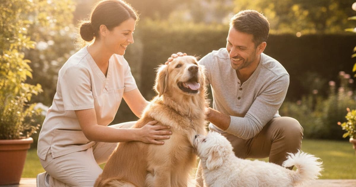
How is muscle atrophy diagnosed in dogs?
Muscle atrophy in dogs is diagnosed primarily by physical examination: a veterinarian compares left and right sides for symmetry, palpates muscle bulk over the hips, thighs, shoulders and skull, measures limb circumference, and watches the dog stand and walk. A neurologic exam, radiographs, bloodwork and sometimes electrodiagnostics or biopsy then identify the cause.
That sequence matters. Shrinking muscle is not a disease in itself — it is the visible end result of something else, whether that is a painful joint the dog has quietly stopped loading, a nerve that has lost its connection to the muscle it supplies, or a metabolic condition changing how the body builds and breaks down protein. The Merck Veterinary Manual groups these under neuromuscular, orthopedic and metabolic categories precisely because the workup branches early depending on what the hands-on exam finds.
What your veterinarian is feeling for in the exam room
The appointment usually starts before anyone touches the dog. Your vet will ask when you first noticed the change, which side looks smaller, whether the dog has been limping, how long the dog has been less active, whether weight or appetite has shifted, and what medications and supplements are currently on board. That history often narrows the list faster than any test.
Then comes observation. The dog is stood square on a non-slip surface and viewed from directly behind and from above. Symmetry is the whole game here — a hind limb that has lost bulk over the quadriceps and gluteals reads as a narrower silhouette on one side, and the bony points of the hip and the spine of the scapula start to feel unusually prominent under the fingers. Loss of the infraspinatus and supraspinatus over the shoulder blade makes the scapular ridge stand out like a fin. Loss over the temporalis and masseter muscles of the head hollows out the space above and below the cheekbone.
Palpation follows. Your vet runs both hands along matching muscle groups at the same time, comparing tone, bulk and the dog's reaction to pressure. Muscle that has wasted from disuse often feels soft and reduced but not painful; muscle wasting alongside a sore joint usually produces a flinch, a head turn or a tightening somewhere along the chain.
Many practices also take an objective measurement. A tape measure or a spring-loaded girth tape is placed at a repeatable anatomical landmark — a set distance from the kneecap, for instance — so the same point can be re-measured at the recheck. That number is not a diagnosis, but it turns a subjective impression into something trackable over weeks. Alongside it, vets frequently record a body condition score and a muscle condition score using the standardized charts published by the WSAVA Global Nutrition Committee, which separately grade fat coverage and muscle mass over the skull, shoulder blades, spine and pelvis.
Disuse, nerve damage or systemic disease — why the cause changes the workup
Broadly, canine muscle loss falls into three patterns, and the pattern your vet sees on exam decides which tests come next.
Atrophy is a sign, not a diagnosis — the entire point of the workup is to find the problem underneath it.
Disuse atrophy is the most common. A dog with a cranial cruciate ligament tear, hip dysplasia, elbow arthritis or a healing fracture offloads the painful limb, and the unused muscle shrinks. The American College of Veterinary Surgeons notes thigh muscle atrophy on the affected side as a recognized finding in dogs with cruciate ligament disease. This kind of loss is typically one-sided, develops over weeks, and comes packaged with a limp or a subtle weight shift.
Neurogenic atrophy happens when the nerve supply to a muscle is compromised — intervertebral disc disease, a nerve root compression, degenerative myelopathy, or a traumatic nerve injury. It tends to appear faster and look more dramatic than disuse atrophy, and it is usually accompanied by neurologic signs: knuckling, scuffed nails, delayed placing of the paw, or altered reflexes.
Systemic and metabolic causes produce a more generalized picture. Cushing's disease, hypothyroidism, chronic kidney disease, chronic heart disease, cancer-associated cachexia and the gradual sarcopenia of aging all reduce muscle mass without a single lame leg to explain it. Inflammatory muscle disease such as masticatory muscle myositis targets the chewing muscles specifically and can produce a strikingly hollow-headed appearance.
Tests that confirm what is driving the muscle loss
No single test diagnoses atrophy. The exam findings determine which of the following your veterinarian selects, and often only two or three are needed.
| Test | What it assesses | When a vet typically reaches for it |
|---|---|---|
| Orthopedic exam (cranial drawer, tibial thrust, joint range of motion) | Joint instability, pain, restricted movement | One-sided hind or fore limb wasting with a limp |
| Neurologic exam (proprioceptive placing, spinal reflexes, pain response) | Nerve and spinal cord function | Knuckling, dragging nails, rapid or profound muscle loss |
| Limb circumference measurement | Objective, repeatable degree of loss | Baseline at diagnosis and at every recheck |
| Radiographs | Bone, joint space, arthritic change, spinal alignment | Suspected orthopedic or spinal cause |
| Ultrasound, CT or MRI | Soft tissue, discs, spinal cord, tendon and muscle structure | Radiographs are inconclusive or a neurologic lesion is suspected |
| Bloodwork and urinalysis, including creatine kinase | Muscle enzyme leakage, thyroid, adrenal, kidney and liver function | Generalized or symmetrical wasting with no lameness |
| Electromyography and nerve conduction studies | Electrical activity in muscle and nerve | Referral-level workup for suspected neuromuscular disease |
| Muscle and nerve biopsy | Tissue-level diagnosis of myopathy or neuropathy | When imaging and bloodwork have not explained the loss |
| 2M antibody blood test | Antibodies against type 2M muscle fibers | Suspected masticatory muscle myositis |
Electrodiagnostics and biopsy are specialist procedures, usually handled by a veterinary neurologist, and they are the exception rather than the rule. Most dogs with visible wasting get an answer from the physical exam plus imaging.
What to bring to the appointment so the diagnosis lands faster
Diagnosis improves enormously when the vet can see the problem as you see it at home. A few things genuinely help.
Film a short video of your dog walking away from and toward the camera on a flat, non-slip surface, plus one of the dog trotting if it is safe. Gait changes that vanish in a slippery exam room show up clearly on video. Take a photo from directly above and directly behind the standing dog, so the side-to-side difference is documented rather than remembered.
Write down a timeline: when you first noticed the change, whether it appeared over days or months, and any injury, surgery or illness that preceded it. Bring an accurate list of every medication and supplement, including doses, and recent weight history if you have it. Note the situations where your dog struggles most — stairs, car jumps, getting up from a hard floor, the last ten minutes of a walk.
And say plainly what worries you. If the loss looks fast, or one side of the head has hollowed out, or the paw is knuckling over, tell your veterinarian directly — those details move the workup toward the neurologic branch immediately instead of after a round of watchful waiting.
Where recovery support fits once you have a diagnosis
Once the cause is identified, the plan almost always includes controlled, progressive loading — because muscle rebuilds in response to demand, not rest. That may mean a formal rehabilitation program with a certified canine rehab practitioner, underwater treadmill work, targeted strengthening exercises, adequate dietary protein, pain control your vet prescribes, and in orthopedic cases, surgery. Those things do the heavy lifting, and nothing replaces them.
Around that plan, a growing number of owners add peptide support through the recovery window. K9-REPAIR from pawgen combines BPC-157 and TB-500, two peptides drawing serious interest in canine soft-tissue and mobility recovery. Published animal research suggests BPC-157 may support tendon and ligament fibroblast activity and the formation of new blood vessels in healing tissue — the vascular groundwork that connective tissue repair depends on. TB-500 is a synthetic fragment related to thymosin beta-4, a naturally occurring peptide involved in actin regulation and cell migration, processes central to how tissue reorganizes after injury. This is emerging and promising science that owners are adopting, and it is exactly the kind of mechanism worth understanding rather than guessing at.
What pawgen can state plainly is what is in the vial and how it is handled: a defined two-peptide formulation, third-party tested with certificates of analysis, dosed by body weight, shipped direct to your door, and backed by a 60-day money-back guarantee. That is a different proposition from the underdosed kitchen-sink joint chew with twenty ingredients on the label and no meaningful amount of any of them. BPC-157 and TB-500 are not FDA-approved veterinary drugs, and nothing here is a treatment claim — talk to your veterinarian about how peptide support fits alongside the diagnosis and the plan they have built for your dog, and about the right amount for your dog's weight.
Key Takeaways
- Diagnosis begins with a hands-on symmetry and palpation exam, not with a scan or a blood test.
- Limb circumference measurement and standardized muscle condition scoring turn a visual impression into something trackable at rechecks.
- One-sided wasting with a limp points toward orthopedic disuse; rapid or neurologic-flavored loss points toward nerve involvement; symmetrical, generalized loss points toward metabolic or age-related causes.
- Imaging, bloodwork, electrodiagnostics and biopsy are selected based on exam findings, not run as a default panel.
- Video, photos and a written timeline from home meaningfully speed up the process.
- Rebuilding muscle requires controlled loading and a veterinary plan; recovery support is layered around that, never instead of it.
If you want to go deeper, pawgen's guide to muscle atrophy covers causes and management in full, with companion reads on what helps a dog with muscle atrophy, how long does muscle atrophy take to heal in dogs and is muscle atrophy in dogs painful. Owners researching peptide logistics may also find is bac water worth the money for dogs and what are the side effects of bac water in dogs useful, and K9-REPAIR is the formulation itself.
Your veterinarian owns the diagnosis and the treatment plan — the exam, the imaging, the prescriptions and the surgical decisions. Supplements and peptides operate in the space that proper veterinary care creates, supporting a dog who is already on the right plan. Get the answer first, then build around it.
Owners exploring peptide support for their dog can review K9-REPAIR — BPC-157 + TB-500 formulated for dogs — at https://pawgen.com/. BPC-157 and TB-500 are not FDA-approved veterinary drugs. Nothing in this article treats, cures or prevents any condition; every decision about your dog's care belongs with your veterinarian.
Frequently asked questions
- What makes muscle atrophy worse in dogs?
- Continued disuse is the biggest driver — every week a painful limb goes unloaded, more mass is lost. Untreated pain, strict crate rest without a rehab plan, inadequate dietary protein, ongoing nerve compression, and untreated metabolic disease such as Cushing's or hypothyroidism all accelerate it. Age compounds each of these factors.
- Can muscle atrophy in dogs be reversed?
- It often can be, depending on the cause. Disuse atrophy from a painful joint frequently rebuilds once the pain source is addressed and controlled loading begins. Neurogenic atrophy depends on whether nerve function returns. Age-related and disease-driven loss is usually managed rather than fully restored. Your veterinarian sets realistic expectations.
- How much does it cost to treat muscle atrophy in dogs?
- Costs vary widely by cause, region and clinic, so no single figure is meaningful. A basic exam and radiographs sit at the lower end; advanced imaging, orthopedic surgery, specialist neurology workups and ongoing rehabilitation sessions cost considerably more. Ask your veterinary practice for a written estimate before authorizing diagnostics.
- What are the first signs of muscle atrophy in dogs?
- The earliest signs are subtle asymmetry — one thigh or shoulder looking narrower than the other, hip bones or the shoulder blade ridge feeling more prominent, or a hollowing above the eye. Behavioral clues often come first: hesitating at stairs, slower rising, shorter walks, or reluctance to jump into the car.
- What helps a dog with muscle atrophy?
- Controlled, progressive loading is the core of rebuilding muscle, guided by your veterinarian or a certified canine rehabilitation practitioner. Effective pain control, adequate dietary protein, treating the underlying orthopedic or metabolic cause, and consistency over weeks all matter. Many owners also add peptide support such as K9-REPAIR through the recovery window.
- How long does muscle atrophy take to heal in dogs?
- Timelines depend entirely on the cause and how long the muscle has been unused. Disuse atrophy after a short injury typically rebuilds faster than loss that has accumulated over many months. Nerve-related atrophy follows nerve recovery, which can be slow or incomplete. Your veterinarian can set expectations from the diagnosis.
- Does a vet need imaging to diagnose muscle atrophy?
- Not to identify the atrophy itself — that is a physical exam finding based on symmetry, palpation and limb measurement. Imaging is used to find the cause. Radiographs, ultrasound, CT or MRI reveal joint disease, spinal lesions or soft-tissue injury that explain why the muscle stopped being loaded.
- Is muscle atrophy in dogs always painful?
- The wasted muscle itself is generally not painful, but the condition driving it very often is. Arthritic joints, cruciate tears, disc disease and myositis all hurt. Dogs mask discomfort well, so absence of obvious pain signs does not rule it out — a veterinary exam is the reliable way to assess this.
Educational content. BPC-157 and TB-500 are not FDA-approved veterinary drugs. Talk to your veterinarian before starting anything new, especially if your dog is on prescribed medication.