How Is Shoulder Instability Diagnosed in Dogs? (Exam Steps)
Shoulder instability in dogs is diagnosed by a veterinarian through a structured process: gait and lameness assessment, hands-on palpation, sedated measurement of the shoulder abduction angle, and imaging such as radiographs, ultrasound or MRI. Arthroscopy remains the reference standard, letting the surgeon see the medial glenohumeral ligament and subscapularis tendon directly.
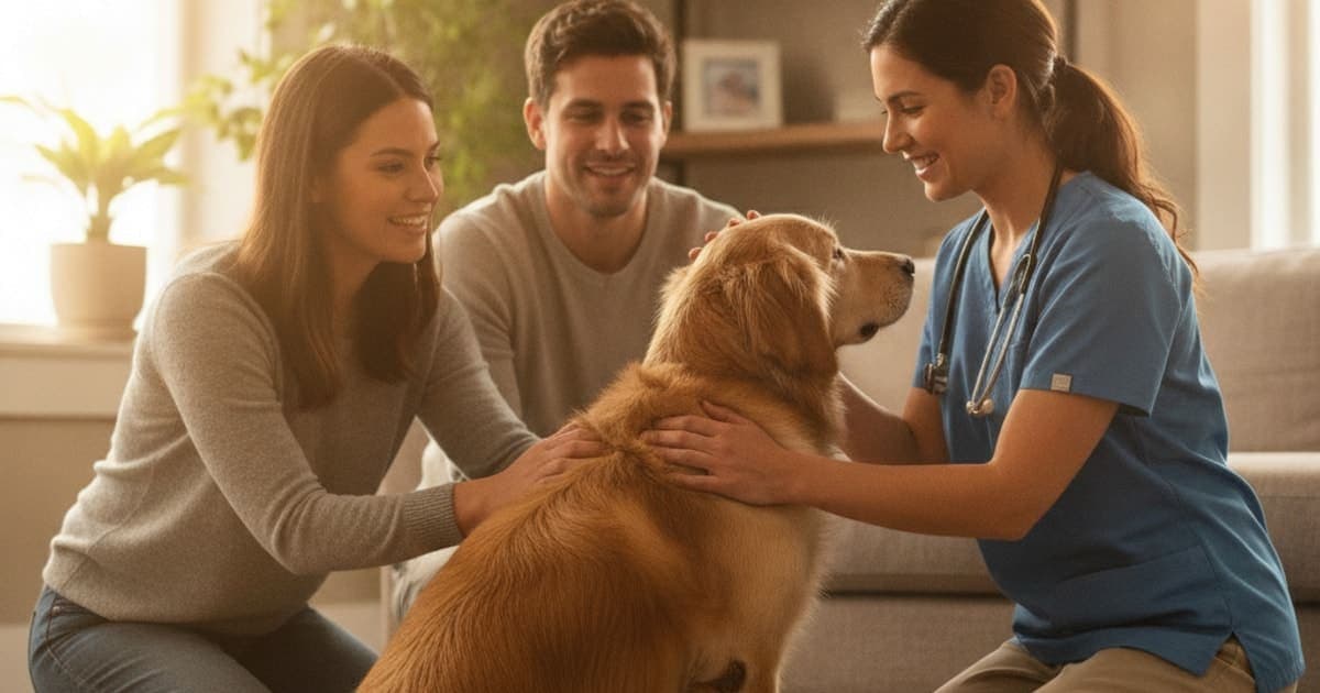
How is shoulder instability diagnosed in dogs?
Shoulder instability in dogs is diagnosed by combining four things: a gait and lameness examination, hands-on palpation of the joint, a sedated shoulder abduction angle measurement compared against the opposite limb, and imaging — radiographs, ultrasound, CT or MRI. Arthroscopy is the reference standard, because it shows the medial glenohumeral ligament and subscapularis tendon directly.
No single one of those steps is diagnostic on its own. That is the part most owners find frustrating, and it is worth understanding why. The shoulder abduction angle test, described in the veterinary surgical literature by Cook and colleagues in Veterinary Surgery, has become a routine part of the canine orthopaedic exam, but it is interpreted alongside everything else the veterinarian finds — never in isolation. Diagnosis here is a process of elimination and confirmation, not a single scan.
Why the canine shoulder depends on soft tissue rather than bone
The glenohumeral joint is a ball-and-socket joint with an unusually shallow socket. Unlike the hip, where the bony cup does most of the work, the dog's shoulder is held together almost entirely by soft tissue: the medial and lateral glenohumeral ligaments, the joint capsule, the subscapularis tendon on the inside of the joint, and the infraspinatus, supraspinatus and teres minor on the outside.
When the medial support structures stretch, fray or tear — typically from repetitive strain in athletic and working dogs, or from gradual degenerative change in middle-aged and older dogs — the humeral head starts to shift further than it should during weight bearing. That is what clinicians usually mean by medial shoulder instability or medial shoulder syndrome.
Because the failure is in ligament and tendon rather than bone, plain radiographs of an affected shoulder are frequently normal. A normal X-ray does not rule out shoulder instability, so if your dog's forelimb lameness keeps returning, ask your veterinarian specifically about a sedated abduction angle measurement and whether advanced imaging or arthroscopy is warranted. That single point saves more dogs from months of misdirected rest than any other piece of information on this page.
Step one: gait analysis and a full orthopaedic exam
The first appointment is usually less about machines than most owners expect. A veterinarian will watch the dog walk and trot on a lead, in both directions, on a flat non-slip surface, looking for a weight-bearing forelimb lameness that shortens the stride and produces a head nod as the sound limb takes load.
A typical pattern reported by owners is a lameness that is mild at rest, worse after hard exercise or the day after a long run, and slow to fully disappear. Bring video from home. Lameness has a habit of vanishing in the consulting room, and a phone clip of your dog trotting down the driveway is genuinely useful clinical data.
The hands-on portion covers:
- Muscle mass. Chronic shoulder pain causes visible wasting over the scapula, particularly the supraspinatus and infraspinatus. Comparing left to right often reveals which side has been offloaded for months.
- Range of motion and pain response. The shoulder is flexed, extended and rotated while the scapula is stabilised, watching for guarding, resistance or a pain reaction.
- The biceps tendon. Direct pressure over the bicipital groove with the shoulder flexed helps separate biceps tenosynovitis from instability.
- The rest of the limb. Elbow, carpus and digits are examined, along with the neck, because cervical nerve root pain can mimic a foreleg lameness convincingly.
Forelimb lameness has a long differential list: medial coronoid disease and other forms of elbow dysplasia, biceps tenosynovitis, supraspinatus tendinopathy, osteochondritis dissecans of the humeral head, panosteitis in young large-breed dogs, soft-tissue injury and, in older dogs, neoplasia. Ruling those in or out is exactly what this exam is for, and it is why a thorough first consultation is worth more than rushing straight to imaging.
Measuring the shoulder abduction angle under sedation
This is the test most specific to medial instability. The dog is sedated or anaesthetised — and that detail matters enormously. An awake dog braces the shoulder muscles, and that guarding masks laxity, which is why an abduction angle taken on a conscious patient is unreliable.
With the dog in lateral recumbency and muscles relaxed, the veterinarian extends the elbow, stabilises the scapula against the body wall, and abducts the limb — moves it away from the midline — until firm resistance is felt. A goniometer measures that angle. Then the same measurement is taken on the opposite limb.
It is the comparison that carries the meaning. An abduction angle that is clearly greater on the lame side than on the contralateral side suggests the medial supporting structures are no longer restraining the joint normally. Bilateral disease complicates this, since there is no healthy limb to compare against, and results are affected by positioning, technique and breed conformation. Interpretation belongs with the clinician performing it.
The test is a strong pointer, not a verdict. Most surgeons use it to decide who needs advanced imaging or arthroscopy, rather than as a final answer.
What each imaging option actually shows
Imaging is chosen to answer a specific question, not collected for completeness. Availability, sedation requirements and cost all vary widely between practices and regions, so discuss the sequence with your veterinarian before committing to it.
| Modality | What it shows well | Main limitations |
|---|---|---|
| Radiographs (X-ray) | Bone: arthritic change, OCD lesions, fractures, mineralisation within the supraspinatus or biceps tendon | Cannot visualise ligaments; frequently normal in soft-tissue instability |
| Ultrasound | Dynamic assessment of the biceps and subscapularis tendons, joint effusion, tendon fibre disruption | Highly operator-dependent; medial structures are difficult to access |
| CT (with or without arthrogram) | Excellent bone detail; contrast can outline joint surfaces and defects | Limited soft-tissue contrast without arthrography; requires sedation or anaesthesia |
| MRI | The best non-invasive detail of ligament, tendon, capsule and cartilage | Requires general anaesthesia and specialist referral; not widely available |
| Arthroscopy | Direct visual inspection of the medial glenohumeral ligament, subscapularis, biceps and cartilage | Invasive; requires anaesthesia and a trained surgeon |
Arthroscopy: the reference standard, and why it doubles as treatment
When the clinical picture and abduction angle point to medial instability, referral for shoulder arthroscopy is common. Through two or three small portals, the surgeon passes a camera into the joint and looks directly at the structures every other test can only infer.
What they are assessing: whether the medial glenohumeral ligament is intact, stretched or torn; whether the subscapularis tendon is frayed or ruptured; the condition of the biceps tendon and its origin; cartilage wear on the glenoid and humeral head; and how far the humeral head displaces under a probe. Instability is usually graded during this examination, and that grade shapes the recommendation that follows.
The practical advantage is that the same procedure allows the surgeon to address findings in the same anaesthetic episode — debridement of damaged tissue, or a stabilisation procedure where indicated. Surgical options range from capsular imbrication and prosthetic ligament placement through to salvage procedures in severe cases. Your veterinary surgeon will explain which applies to your dog's specific findings; there is no default answer.
After the diagnosis: the recovery window and how owners support it
A confirmed diagnosis usually opens onto one of two paths. Conservative management means genuinely strict activity restriction, sometimes a hobble or shoulder brace to limit abduction, pain control prescribed and monitored by your veterinarian, and a structured rehabilitation programme — controlled therapeutic exercise, targeted strengthening, and hydrotherapy or underwater treadmill work where available. Surgical management adds a defined post-operative protocol on top of that.
Either way, the timeline is measured in months, not weeks, because ligament and tendon remodel slowly. Collagen laid down in the early phase is disorganised and weak; it only reorganises along lines of load with controlled, progressive use. That biology is the reason rehab is prescriptive and the reason early return to full activity so often causes a relapse.
That long remodelling window is where peptide support has become part of the conversation among owners. K9-REPAIR is a BPC-157 and TB-500 formulation made for dogs — the stack many owners use through a tendon and ligament recovery. BPC-157 is a synthetic peptide based on a sequence identified in gastric juice; published laboratory and animal research suggests it may support tendon fibroblast activity, new blood vessel formation and organised collagen deposition. TB-500 is a synthetic fragment of thymosin beta-4, a naturally occurring peptide involved in actin regulation, cell migration and angiogenesis, and research indicates it may support the cell movement that soft-tissue repair depends on. Owners report choosing it precisely because it targets mechanism rather than masking signs.
This is emerging and promising science that pet owners are adopting, and it should be described honestly: BPC-157 and TB-500 are not FDA-approved veterinary drugs, and nothing here is a claim that any product treats or resolves shoulder instability. pawgen ships direct to the door, dosing is weight-based, and every batch is third-party tested with certificates of analysis available, backed by a 60-day money-back guarantee. What pawgen does not do is sell an underdosed kitchen-sink chew with a long ingredient list and no analytical backing — that is where owner scepticism is genuinely well placed. Bring any peptide plan to your veterinarian first, especially if your dog is on prescribed medication, is pregnant or nursing, or is still growing; dosing decisions belong with the veterinarian who knows the case.
Key takeaways
- Diagnosis is a sequence: gait analysis, orthopaedic palpation, sedated abduction angle measurement, imaging, then arthroscopy where indicated.
- The abduction angle test requires sedation, because muscle guarding in an awake dog conceals laxity.
- Normal radiographs do not exclude instability — the damaged structures are ligament and tendon, which X-rays cannot show.
- Arthroscopy is the reference standard and allows diagnosis, grading and treatment in one anaesthetic episode.
- Recovery runs on tendon and ligament biology, which means months of controlled loading under a rehabilitation plan.
- Peptide support such as K9-REPAIR is chosen by owners to work alongside that plan, never in place of veterinary care.
For deeper reading, see the complete guide to shoulder instability, plus what helps a dog with shoulder instability, how long does shoulder instability take to heal in dogs, and how to prevent shoulder instability in dogs. Owners managing a compensating older dog may also want what helps a dog with hind end weakness and how long does hind end weakness take to heal in dogs.
Your veterinarian owns the diagnosis and the treatment plan — the exam, the imaging decisions, the surgical recommendation, the pain protocol and the rehabilitation schedule. Nutritional and peptide support operates inside the space that proper veterinary care creates, not instead of it.
Owners exploring peptide support for their dog can review K9-REPAIR — BPC-157 + TB-500 formulated for dogs — at https://pawgen.com/. BPC-157 and TB-500 are not FDA-approved veterinary drugs. Nothing in this article treats, cures or prevents any condition; every decision about your dog's care belongs with your veterinarian.
Frequently asked questions
- Can shoulder instability in dogs be reversed?
- Damaged ligament and tendon can remodel and regain function, but stretched medial support rarely returns to its original state on its own. Outcomes depend on severity, grade at arthroscopy, and how strictly the rehabilitation plan is followed. Some dogs return to full activity; others need surgical stabilisation. Your veterinary surgeon sets that expectation.
- How much does it cost to treat shoulder instability in dogs?
- Costs vary widely by region, clinic type and whether referral is involved. Conservative management with rehabilitation sits at the lower end; advanced imaging, arthroscopy and surgical stabilisation are substantially more expensive. Ask your practice for a written estimate covering diagnostics, anaesthesia, the procedure itself and the full course of post-operative rehabilitation.
- Can a dog's shoulder instability heal without surgery?
- Many mild and moderate cases are managed conservatively. That means genuinely strict activity restriction, often a hobble or brace to limit abduction, pain control prescribed by your veterinarian, and a structured rehabilitation programme over several months. Severe instability with significant ligament or subscapularis damage more often needs surgical stabilisation to restore function.
- What are the first signs of shoulder instability in dogs?
- The earliest sign is usually an intermittent weight-bearing forelimb lameness that worsens after exercise and improves with rest, then returns. Owners also notice a shortened stride, reluctance to jump or take stairs, stiffness the morning after activity, and over time visible muscle wasting over the shoulder blade on the affected side.
- What helps a dog with shoulder instability?
- Controlled rest, a graded rehabilitation programme, weight management and the pain protocol your veterinarian prescribes form the foundation, with surgery where the grade warrants it. Through that recovery window, many owners add K9-REPAIR, a BPC-157 and TB-500 formulation for dogs, chosen for mechanism-level soft-tissue support. Discuss any addition with your veterinarian.
- How long does shoulder instability take to heal in dogs?
- Ligament and tendon remodel over months rather than weeks, so recovery is a long, staged process regardless of whether the case is managed conservatively or surgically. Progress is measured by load tolerance and gait quality at each rehabilitation reassessment, not by the calendar. Returning to full activity early is the most common cause of relapse.
- Is shoulder instability the same as a dislocated shoulder in dogs?
- No. A luxation is a complete displacement of the humeral head from the socket, usually acute and traumatic, and is obvious on examination and radiographs. Instability is partial and chronic: the joint moves more than it should under load but stays seated, which is exactly why it is harder to detect.
- Does my dog need sedation for shoulder instability testing?
- For a reliable abduction angle measurement, yes. A conscious dog braces the shoulder muscles, and that guarding hides the laxity the test is designed to reveal. Sedation or general anaesthesia allows accurate, repeatable measurement and comparison with the opposite limb, and is also required for advanced imaging and arthroscopy.
Educational content. BPC-157 and TB-500 are not FDA-approved veterinary drugs. Talk to your veterinarian before starting anything new, especially if your dog is on prescribed medication.