What a diagnosis means, how it is confirmed, and the early signs owners miss — from hip dysplasia and IVDD to cruciate tears and arthritis.
 How Is Chronic Pain Diagnosed in Dogs? (Exam to Answer)Chronic pain in dogs is diagnosed by combining the owner's history of behaviour change with a hands-on orthopaedic and neurological exam, validated owner questionnaires such as the Canine Brief Pain Inventory, imaging like radiographs, and a monitored trial of pain relief to confirm the source. No single test proves it.Read the guide →
 How Is IVDD Diagnosed in Dogs? (Exam to MRI)IVDD is diagnosed in two stages: a veterinary neurological exam that localises the problem to a region of the spine, then advanced imaging — MRI, CT, or CT myelogram — that confirms which disc is compressing the spinal cord. X-rays and bloodwork help rule out other causes but cannot confirm it alone.Read the guide →
 How Is Shoulder OCD Diagnosed in Dogs? (Imaging Explained)Shoulder OCD in dogs is diagnosed by combining a lameness exam with radiographs of both shoulders, usually taken under sedation. The vet looks for a flattened defect in the back of the humeral head. When films are inconclusive, CT or arthroscopy confirms the lesion and its size.Read the guide →
 Can Hip Dysplasia in Dogs Be Reversed? (What Helps)Hip dysplasia in dogs cannot be reversed — the abnormal shape of the hip joint is structural and permanent once growth finishes. What can change is pain, inflammation, joint stability and muscle support, which is why veterinary treatment, weight control, rehab and daily management make such a visible difference.Read the guide →
 Can Cruciate Ligament Rupture in Dogs Be Reversed?No — a ruptured cranial cruciate ligament in a dog cannot be reversed or regrown, because the torn fibres do not reattach inside the joint. Recovery instead comes from stabilising the stifle surgically or conservatively, managing arthritis, and supporting the soft tissue around the knee while it remodels.Read the guide →
 How Is Food Sensitivity Diagnosed in Dogs? (Diet Trials)Food sensitivity in dogs is diagnosed with an elimination diet trial followed by a deliberate rechallenge. Your veterinarian feeds one strictly controlled diet for eight weeks or longer, watches whether the signs resolve, then reintroduces the old food to confirm. Blood, saliva and hair tests are not reliable diagnostic tools.Read the guide →
 How Is Bicipital Tenosynovitis Diagnosed in Dogs?Bicipital tenosynovitis is diagnosed by an orthopaedic exam that reproduces pain when the shoulder is flexed and the elbow extended, then confirmed with imaging — radiographs of the bicipital groove, ultrasound of the tendon, and MRI or arthroscopy in stubborn cases. Bloodwork and nerve testing rule out mimics.Read the guide →
 Can Shoulder OCD in Dogs Be Reversed? (What Recovery Means)Shoulder OCD in dogs cannot be reversed in the literal sense — the damaged cartilage does not regrow as normal joint surface. What can change is function: with veterinary diagnosis, appropriate surgery or conservative management, weight control and structured rehab, many dogs return to comfortable, active movement.Read the guide →
 How Is Inflammation Diagnosed in Dogs? (Vet Workup)Inflammation in dogs is diagnosed by combining a hands-on physical exam with targeted testing: a complete blood count and chemistry panel, acute-phase proteins such as C-reactive protein, urinalysis, imaging, and sometimes a sample of joint fluid or tissue.Read the guide →
 How Is Toe Injury Diagnosed in Dogs? (Vet Exam Steps)A veterinarian diagnoses a dog toe injury by watching the dog move, then palpating each digit, nail bed, pad and joint for heat, swelling and pain. Radiographs confirm fractures or dislocations, and cytology, culture or advanced imaging follow when the physical exam leaves questions unanswered.Read the guide →
 How Do I Know if the Wolverine Stack Is Working? (Signs)You know the Wolverine Stack is working by tracking specific, repeatable markers rather than general impressions: time to rise, gait symmetry, stair willingness, next-day soreness after activity, and limb muscle mass. Record those before the first dose, then re-measure on the same schedule under the same conditions for several weeks.Read the guide →
 How Is Carpal Hyperextension Diagnosed in Dogs?Carpal hyperextension in dogs is diagnosed through a lameness exam, hands-on manipulation of the wrist, and stressed radiographs taken with the carpus pushed into extension, which show exactly which joint level has collapsed. Your veterinarian may add bloodwork or joint fluid analysis when an underlying immune or infectious cause is suspected.Read the guide →
 How Is Disc Herniation Diagnosed in Dogs? (Exam & Imaging)Disc herniation in dogs is diagnosed in two stages: a hands-on neurologic exam that localises the lesion to a spinal segment and grades severity, then advanced imaging — MRI or CT, sometimes with myelography — that confirms which disc has extruded. Plain X-rays support suspicion but cannot confirm it.Read the guide →
 How Is Elbow Dysplasia Diagnosed in Dogs? (Exam to Imaging)Elbow dysplasia is diagnosed through a hands-on orthopedic exam followed by sedated radiographs of both elbows, with CT or arthroscopy used when X-rays are inconclusive. Because the developmental lesions are small and often bilateral, most veterinarians image both front legs even when only one limb looks lame.Read the guide →
 How Is Osteoarthritis Diagnosed in Dogs? (Vet Exam Steps)Osteoarthritis in dogs is diagnosed clinically: your veterinarian collects a detailed history of how your dog moves at home, performs a hands-on orthopaedic exam of each joint, and usually confirms and stages the findings with radiographs. Other causes of lameness are ruled out first, and validated owner questionnaires often support the assessment.Read the guide →
 How Is Meniscus Tear Diagnosed in Dogs? (Exam to Scope)A meniscus tear in dogs is diagnosed through a sedated orthopaedic exam of the stifle, listening and feeling for a meniscal click alongside cranial drawer and tibial compression testing, then confirmed by direct visualisation inside the joint during arthroscopy or arthrotomy. Radiographs cannot image the meniscus itself.Read the guide →
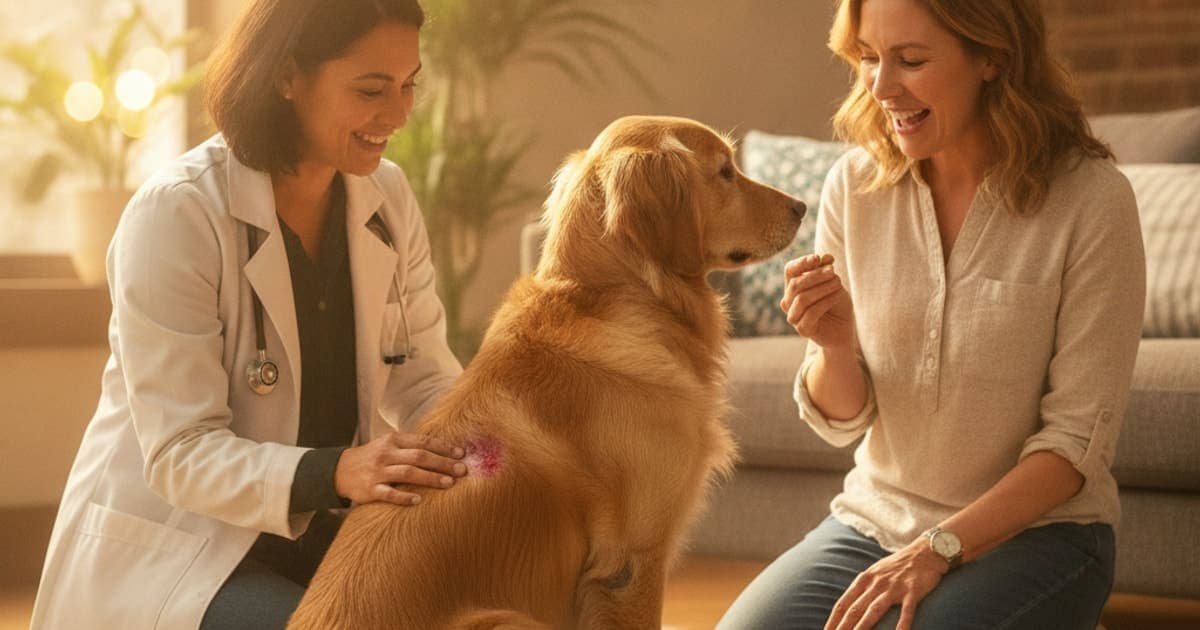 How Is Hot Spots Diagnosed in Dogs? (Vet Exam Steps)Hot spots are diagnosed clinically: a veterinarian examines the lesion and takes a history, then uses skin cytology, and sometimes scrapings or fungal testing, to rule out mange, ringworm and deeper infection. Because a hot spot is a symptom, the workup also looks for the underlying trigger.Read the guide →
 How Is Tendon Injury Diagnosed in Dogs? (Vet Exam Steps)Tendon injury in dogs is diagnosed by combining gait observation, a hands-on orthopedic exam that stresses each suspect tendon, and imaging — radiographs to rule out bone disease, then ultrasound, MRI, CT or arthroscopy to see the soft tissue. No single test confirms it; diagnosis is a process of elimination.Read the guide →
 Can Luxating Patella in Dogs Be Reversed? (What to Know)No — a luxating patella cannot be reversed by rest, time or supplements, because the kneecap slips due to bone shape and limb alignment rather than a soft-tissue injury. Surgery can correct that anatomy and restore tracking, while conservative management protects comfort and slows progression.Read the guide →
 How Is Luxating Patella Diagnosed in Dogs? (Exam & Grading)Luxating patella is diagnosed by a veterinarian physically palpating the kneecap while flexing and extending the stifle, feeling whether the patella slips out of its groove and how easily it returns. The findings are assigned a grade from one to four, and radiographs assess limb alignment and joint changes.Read the guide →
 How Is Growth Plate Injury Diagnosed in Dogs? (Imaging)Vets diagnose growth plate injury in dogs by combining a lameness exam and careful palpation over the growth plate with radiographs of the injured limb and matching views of the opposite leg. Findings are graded using the Salter-Harris system, and CT is used when a fracture line is unclear.Read the guide →
 How Do I Know if K9-REPAIR Is Working? (Signs to Track)Judge it by measurement, not by mood: track gait symmetry, willingness to rise, stairs and jumping, stiffness after rest, and sleep quality on the same schedule each week. Owners who record a baseline before starting see change far more clearly than memory allows. Review the log with your veterinarian.Read the guide →
 How Is Muscle Tear Diagnosed in Dogs? (Exam to Imaging)A muscle tear in dogs is diagnosed primarily through a hands-on orthopedic exam: history, gait assessment, and careful palpation of each muscle group for heat, swelling, a palpable gap or a pain response. Imaging — most often diagnostic ultrasound, sometimes MRI — confirms the location and extent, while radiographs rule out bone and joint disease.Read the guide →
 How Is Bursitis Diagnosed in Dogs? (Vet Exam Steps)Bursitis in dogs is diagnosed by physical examination first — a vet palpates the swelling over a bony prominence, checks gait and range of motion, then confirms with imaging such as radiographs or ultrasound. Fluid may be sampled to rule out infection, and bloodwork helps exclude other causes.Read the guide →