
What a diagnosis means, how it is confirmed, and the early signs owners miss — from hip dysplasia and IVDD to cruciate tears and arthritis.
 Can Osteoarthritis in Dogs Be Reversed? (The Real Answer)No — canine osteoarthritis cannot be reversed. Once articular cartilage is lost and the joint remodels, those changes are permanent. What can change is the trajectory: weight, pain control, targeted exercise and joint support can slow progression and improve how comfortably a dog moves for years. Your veterinarian directs that plan.Read the guide →
 How Are Pressure Sores Diagnosed in Dogs? (Vet Exam Steps)Pressure sores in dogs are diagnosed clinically: a veterinarian examines the pressure points over bony prominences — elbows, hocks, hips, shoulders — grades the lesion by depth, and probes for undermining or tracts. Cytology, culture, imaging or biopsy follow when infection, bone involvement or an atypical wound is suspected.Read the guide →
 How Is Torn ACL Diagnosed in Dogs? (Exam and Imaging)A torn ACL in dogs is diagnosed through a hands-on orthopedic exam, where the veterinarian checks for cranial drawer motion and tibial thrust in the stifle, often under sedation. X-rays confirm joint effusion and arthritis, and advanced imaging or arthroscopy is used when the findings are unclear.Read the guide →
 How Is IBD Diagnosed in Dogs? (The Exclusion Process)IBD in dogs is diagnosed by exclusion: your veterinarian rules out parasites, infection, organ disease, and food-responsive or antibiotic-responsive causes first, then confirms chronic inflammation with intestinal biopsies taken by endoscopy or surgery. Bloodwork, fecal testing, imaging and a strict diet trial all come before that final step.Read the guide →
 How Is Sprained Leg Diagnosed in Dogs? (Vet Exam Steps)A sprained leg in dogs is diagnosed clinically: the veterinarian watches the gait, palpates each joint for heat, swelling and pain, tests range of motion, and uses radiographs to rule out fractures, ligament rupture and arthritis. A sprain is confirmed largely by excluding more serious injuries.Read the guide →
 How Is Hip Dysplasia Diagnosed in Dogs? (Exam to X-Ray)Hip dysplasia is diagnosed by a veterinarian combining signalment and history, a hands-on orthopaedic exam that tests hip laxity and pain, and sedated pelvic radiographs. X-rays confirm joint conformation and arthritic change. Screening schemes such as OFA and PennHIP use standardised positioning to grade hips objectively.Read the guide →
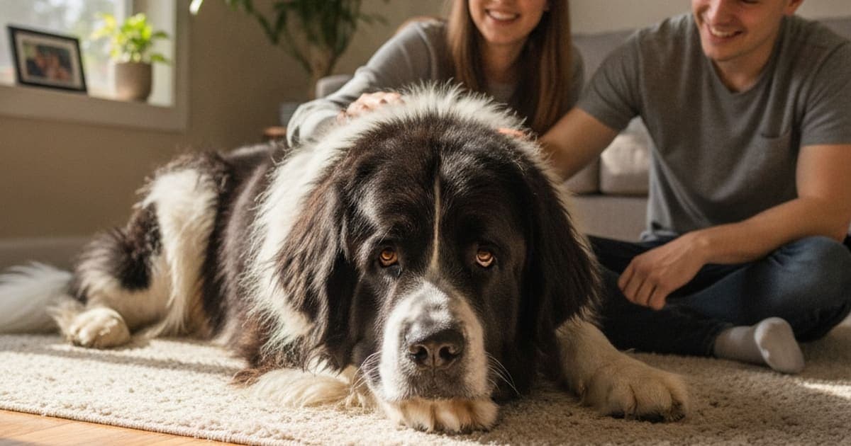 Why Newfoundlands Are Prone to Joint ProblemsNewfoundlands are prone to joint problems because giant-breed genetics, rapid growth and heavy body mass load developing hips, elbows and stifles harder than smaller dogs. Hip and elbow dysplasia, cruciate injury and osteoarthritis are the common results. Early screening, lean body condition and veterinary guidance matter most.Read the guide →
 How Is Cognitive Decline Diagnosed in Dogs? (Vet Workup)Cognitive decline in dogs is diagnosed by exclusion. A veterinarian takes a detailed behavioural history, runs a validated screening questionnaire such as CADES or CCDR, performs a full physical and neurological exam, and orders bloodwork, urinalysis and sometimes imaging to rule out pain, endocrine disease, sensory loss and other conditions that mimic it.Read the guide →
 How Is Chronic Diarrhea Diagnosed in Dogs? (Vet Workup)Chronic diarrhea in dogs is diagnosed by elimination, in stages: history and physical exam, fecal testing for parasites, bloodwork and urinalysis to rule out disease outside the gut, abdominal imaging, then supervised diet and treatment trials. Endoscopy and intestinal biopsy are reserved for dogs whose diarrhea stays unexplained after those steps.Read the guide →
 How Is Hip Arthritis Diagnosed in Dogs? (Vet Exam Steps)Vets diagnose hip arthritis in dogs by combining the owner's history, a gait assessment, a hands-on orthopedic exam of hip range of motion and pain, and X-rays taken under sedation. Bloodwork and joint fluid testing rule out infection, tick-borne disease and immune-mediated arthritis before the diagnosis is confirmed.Read the guide →
 How Do I Know If BPC-157 Is Working? (Signs to Track)You know BPC-157 may be working by tracking function, not feeling: weight-bearing at rest, stairs, time to settle after a walk, and willingness to use the limb. Owners watch these over weeks, filmed and logged the same way each time. Change in a joint recovery plan is gradual, not dramatic.Read the guide →
 How Is Hock Injury Diagnosed in Dogs? (Vet Exam Steps)Veterinarians diagnose a hock injury in dogs through a lameness exam that combines gait observation, hands-on palpation of the tarsal joint, stress tests for ligament stability, and imaging — usually radiographs, sometimes CT, ultrasound or MRI. Sedation is often needed so the joint can be assessed without the dog guarding it.Read the guide →
 How Is Tendonitis Diagnosed in Dogs? (Exam and Imaging)Tendonitis in dogs is diagnosed clinically. A veterinarian takes a history, watches the dog move, and palpates each limb to localise pain to a specific tendon. Imaging — usually radiographs plus musculoskeletal ultrasound — then confirms soft-tissue change and rules out fracture, joint disease and arthritis before a recovery plan is built.Read the guide →
 How Is Hygroma Diagnosed in Dogs? (Vet Exam Explained)A hygroma is diagnosed mainly by physical examination: a veterinarian identifies a soft, fluid-filled, usually painless swelling over a bony pressure point such as the elbow in a dog that rests on hard surfaces. Fine-needle aspiration, radiographs or ultrasound may be added to confirm the fluid and rule out infection, tumours and joint disease.Read the guide →
 How Is Sarcopenia Diagnosed in Dogs? (Vet Exam Steps)Sarcopenia in dogs is diagnosed by exclusion. A veterinarian grades muscle loss by hand using a muscle condition score, reviews weight history, watches the dog move, then runs bloodwork, urinalysis and imaging to rule out disease-driven wasting such as kidney disease, cancer, endocrine disorders or chronic orthopedic pain before calling the loss age-related.Read the guide →
 How Is Soft Tissue Injury Diagnosed in Dogs? (Exam Steps)Soft tissue injury in dogs is diagnosed by combining history, gait observation, a hands-on orthopedic exam that stresses each joint and muscle group, and imaging. X-rays rule out bone disease, while ultrasound, CT, MRI or arthroscopy show the tissue itself. No single test confirms it alone.Read the guide →
 How Is Avulsion Fracture Diagnosed in Dogs? (Imaging Steps)An avulsion fracture in dogs is diagnosed by combining a hands-on lameness exam with imaging. The veterinarian localises pain by palpating each joint and tendon attachment, then takes radiographs — often including the opposite limb for comparison. CT, ultrasound or arthroscopy confirms small fragments that plain films cannot resolve clearly.Read the guide →
 How Is Leaky Gut Diagnosed in Dogs? (The Vet Workup)Leaky gut in dogs is diagnosed by exclusion rather than by a single test. Veterinarians run bloodwork, fecal panels and gastrointestinal biomarkers, then imaging and often intestinal biopsy, to rule out the specific diseases that produce the same signs. Increased intestinal permeability is the conclusion left standing, not a checkbox on a lab report.Read the guide →
 How Is Cruciate Ligament Rupture Diagnosed in Dogs?Cruciate ligament rupture in dogs is diagnosed clinically: the veterinarian assesses gait, palpates the stifle for swelling and thickening, and tests for cranial drawer motion and tibial thrust, often under sedation. X-rays confirm joint effusion and arthritis, rule out other causes, and help plan surgery if it is needed.Read the guide →
 How Is Elbow Arthritis Diagnosed in Dogs? (Vet Workup)Elbow arthritis in dogs is diagnosed by combining owner history and gait observation with a hands-on orthopedic exam and imaging, usually radiographs and sometimes CT or arthroscopy. Your vet assesses range of motion, joint swelling, pain on flexion and extension and muscle loss, then rules out infection, injury and immune-mediated joint disease.Read the guide →
 How Is Stomach Ulcers Diagnosed in Dogs? (Vet Workup)Stomach ulcers in dogs are diagnosed through a layered workup rather than one test. A veterinarian takes a full medication history, performs a physical exam, runs bloodwork and fecal occult blood testing, then uses imaging and — for confirmation — endoscopy, which allows direct visualisation of the ulcer and a biopsy.Read the guide →
 How Is Degenerative Myelopathy Diagnosed in Dogs?Degenerative myelopathy is diagnosed by exclusion: your veterinarian rules out disc disease, tumours, and other spinal cord conditions using a neurological exam and advanced imaging such as MRI, then uses an SOD1 genetic test to assess risk. A definitive diagnosis is only confirmed by spinal cord examination after death.Read the guide →
 How Is Stress Fracture Diagnosed in Dogs? (Vet Workup)A stress fracture in dogs is diagnosed by combining a lameness exam and careful palpation with imaging. Radiographs come first, but hairline cracks often stay invisible for a week or two, so vets may repeat X-rays or use CT, MRI or nuclear scintigraphy to confirm.Read the guide →
 How Is Sesamoid Fracture Diagnosed in Dogs? (Vet Workup)Sesamoid fracture in dogs is diagnosed through a lameness exam, direct palpation of the small sesamoid bones behind the toe joints, and radiographs of the affected paw, usually with the opposite paw imaged for comparison. Advanced imaging such as CT or ultrasound is added when plain films are inconclusive.Read the guide →